Case Author(s): Tate Allen, M.D. and Farrokh Dehdashti, M.D. , 7/29/98 . Rating: #D2, #Q4
Diagnosis: Bilateral Spondylolysis at L5
Brief history:
Low back pain
Images:
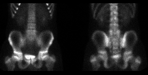
Delayed bone scintigraphy, anterior and posterior images
View main image(bs) in a separate image viewer
View second image(bs).
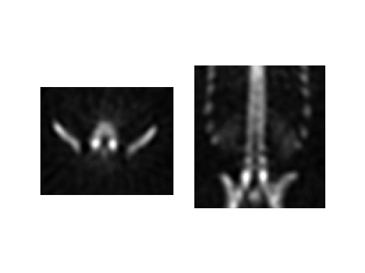
SPECT axial and coronal images at L5
View third image(xr).
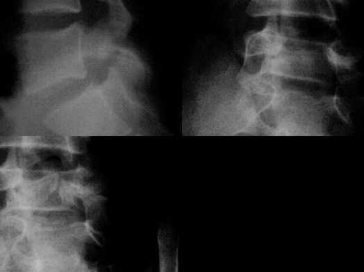
Lateral and bilateral oblique radiolgraphs at L5
Full history/Diagnosis is available below
Diagnosis: Bilateral Spondylolysis at L5
Full history:
A 13-year-old male presents with low back pain exacerbated by physical
activity.
Radiopharmaceutical:
17.9 mCi Tc-99m MDP i.v.
Findings:
Focal increased activity is present within
the posterior elements bilaterally at L5 on both the planar
and SPECT images. These abnormalities correspond with
bilateral pars interarticularis defects seen at L5 on
lumbar radiographs.
Discussion:
Spondylolysis is a defect in the pars interarticularis commonly found
in the lumbar spine at L4 and L5. Frequently, the etiology for this
defect is a stress fracture, although, there may be a congenital
weakness in the osseous matrix of pars interarticularis that predisposes
some individuals to the development of spondylolysis. Individuals
commonly affected are male athletes presenting with back and radicular
pain, however, some patients are asymptomatic. Spondylothisthesis or
slippage of a vertebral body onto another can be associated.
Spondylolysis is usually diagnosed with radiographs of the
lumbar spine. Oblique views which show the "scottie dog" are helpful.
Bone scintigraphy shows increased uptake at the pars defect. In the
early stages of spondylolysis, bone scintigraphy demonstrates a poorly
defined, slightly increased activity that occurs when rapid osteoclastic
resorption exceeds the osteoblastic response). In addition, contralateral
uptake in a unilateral pars defect probably represents a physiological
response or stress fracture in the presence of an unstable neural arch.
The intensity of uptake depends upon the degree of bone repair. Absence
of uptake can be seen in old or stabilized, nonsymptomatic pars defects.
SPECT is superior to planar imaging for detection and accurately
localizing vertebral arch abnormalities.
Reference: Resnick D: Bone and Joint Imaging. Philadephia, WB Saunders
Co, 1996.
Major teaching point(s):
SPECT is especially useful in evaluation of lumbar spine because it
allows for precise localization of a lesion to the vertebral body, disc
space , or vertebral arch.
ACR Codes and Keywords:
- General ACR code: 34
- Spine and Contents:
3.4231 "Spondylolysis without spondylolisthesis"
References and General Discussion of Bone Scintigraphy (Anatomic field:Spine and Contents, Category:Effect of Trauma)
Search for similar cases.
Edit this case
Add comments about this case
Read comments about this case
Return to the Teaching File home page.
Case number: bs094
Copyright by Wash U MO