Case Author(s): Matt Jaksha, M.D. and Henry Royal,M.D. , . Rating: #D2, #Q3
Diagnosis: Small cell carcinoma
Brief history:
66 year old female. Rule out metastases.
Images:
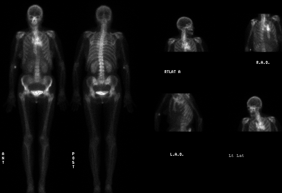
Delayed whole body and spot images
View main image(bs) in a separate image viewer
Full history/Diagnosis is available below
Diagnosis: Small cell carcinoma
Full history:
This 66 year old woman had a known lung cancer, proven by biopsy to be small cell carcinoma. She was being evaluated for metastatice disease to plan therapy.
Radiopharmaceutical:
21.5 mCi Tc-99m MDP i.v.
Findings:
There is abnormal activity in the chest, at the midline and slightly more prominent to the left of midline. The activity is more prominent on the anterior than on the posterior view. On oblique views, it appears to be deep to the sternum.
The skeleton itself is normal. There is no evidence of osseous metastases.
Discussion:
Soft tissue uptake of Tc-99m MDP can have many different causes. Soft tissue activity is often present in conjunction with soft tissue calcification. It can be seen in infarcted tissue. Some tumors show activity on bone scintigraphy. Besides osteogenic sarcomas, mucinous adenocarcinomas (particularly of the breast, colon, and ovary) and neuroblastomas often show activity. Uptake of the agent may be due to gross or microscopic calcification, but the nature of the tumor vascularity and interstitium may also play a role.
Followup:
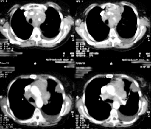
Chest CT demonstrates diffuse soft tissue infiltration of the superior mediastinum, surrounding the aortic arch and great vessels. A 4 x 3 cm mass is seen peripherally in the left upper lobe, maybe pleurally based.There is a moderate left pleural effusion.
View followup image(ct).
Select images from CT of the chest with i.v. contrast
Major teaching point(s):
The differential for uptake of bone agent in the soft tissues includes neoplasm.
(See Heck LL: Gamuts: Exra-osseous localization of phosphate bone agents. Semin Nucl Med 10(3):311-312,1980)
ACR Codes and Keywords:
References and General Discussion of Bone Scintigraphy (Anatomic field:Lung, Mediastinum, and Pleura, Category:Neoplasm, Neoplastic-like condition)
Search for similar cases.
Edit this case
Add comments about this case
Read comments about this case
Return to the Teaching File home page.
Case number: bs091
Copyright by Wash U MO