Case Author(s): Matt Jaksha,M.D. and Keith Fischer, M.D. , 5/21/97
05/21/97 . Rating: #D3, #Q4
Diagnosis: Meningococcemia
Brief history:
9 month old male; rule out osteomyelitis.
Images:

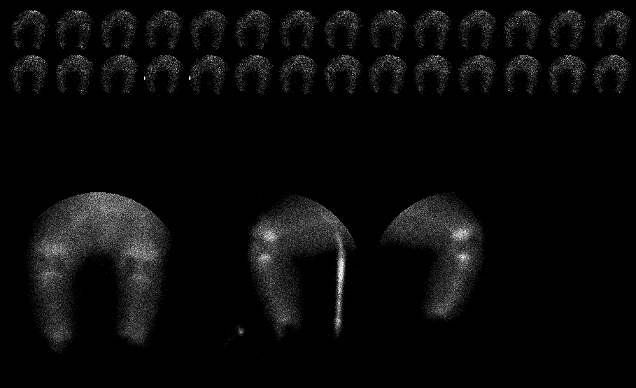
Flow and immediate images of the lower extremities are shown
View main image(bs) in a separate image viewer
View second image(bs).
2 hour delayed images
View third image(bs).
2 hour delayed spot images with increased intensity
Full history/Diagnosis is available below
Diagnosis: Meningococcemia
Full history:
This is a 9 month old male with known meningococcemia. The patient has
had skin grafts to several areas, including medially over the right knee.
There is now concern for infection at that site because of erythema
and drainage. The study is requested to rule out osteomyelitis of the
distal right femur.
Radiopharmaceutical:
Tc-99m MDP
Findings:
The flow and immediate images show no increased activity in the region
of the right knee. However, there is absent activity indicating no
blood flow to the feet and ankles bilaterally. The delayed images
show no activity to either hand. There is increased activity adjacent to theo the
bones of the right forearm, and to a lesser extent at the distal left
forearm.
Note that there is no evidence of osteomyelitis in the region of the right knee.
Discussion:
Neisseria meningitidis (gram negative cocci) is found in the human oropharynx.
It can cause a variety of diseases, including meningitis and bacteremia.
The bacteremia can be associated with purpura and visceral hemorrhages due to endotoxins.
Shock and DIC can develop. Petechiae and areas of purpura can progess
rapidly and be extensive, related to hemorrhage into the skin. Forty to sixty
percent of patients with fulminant meningococcemia die of cardiac/respiratory
collapse. Those who recover may have extensive sloughing of skin lesions and require
amputations for gangrene.
References: Harrison's Principles of Internal Medicine, 12th edition,
pp. 591-592.
Robbins Pathologic Basis of Disease, 4th edition, p. 1253.
Followup:
Amputations were planned for this child because of the obvious gangrene
involving hands and feet. Radiographs of the extremities demonstrated
extensive soft tissue swelling and loss of normal fascial planes. The
right forearm shows frank calcification within the soft tissues related
to the necrosis (see followup image).
View followup image(xr).
AP and lateral view of the right forearm
Major teaching point(s):
Delivery of radiopharmaceuticals to an area is dependent upon blood flow
to that area.
Uptake of Tc-99m MDP in the soft tissues can be related to calcium in the
tissue, and may be seen before the calcification is identified radiographically.
ACR Codes and Keywords:
References and General Discussion of Bone Scintigraphy (Anatomic field:Vascular and Lymphatic Systems, Category:Inflammation,Infection)
Search for similar cases.
Edit this case
Add comments about this case
Return to the Teaching File home page.
Case number: bs090
Copyright by Wash U MO

