Case Author(s): M. Quinn, MD and M. Mintun, MD , 01/30/98 . Rating: #D2, #Q4
Diagnosis: GU-GI fistula
Brief history:
61 yo male with back pain
Images:
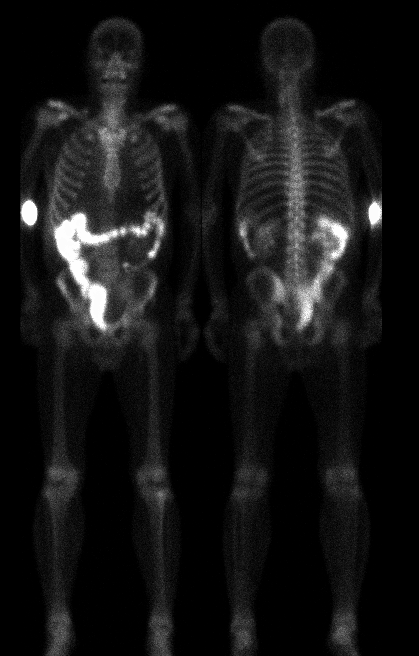
Anterior and posterior images are shown
View main image(bs) in a separate image viewer
Full history/Diagnosis is available below
Diagnosis: GU-GI fistula
Full history:
61 year old male with prior cystectomy and diverting colostomy for
transitional cell carcinoma of the bladder with colonic invasion and
obstruction. Patient had a surgically created neobladder formed from
a large segment of ileum. He recently was experiencing back pain and
bone scan was done to exclude osseous metastatic disease.
Radiopharmaceutical:
Tc99m-MDP
Findings:
There is no evidence for osseous metastases. However, there is abnormal
radiopharmaceutical accumulation throughout the large bowel from the
right colon to the site of the diverting colostomy.
Discussion:
Obviously, the presence of radiopharmaceutical in the colon is abnormal
on delayed images from a bone scan. This appearance can be caused by
previous administration of other imaging agents normally excreted
through the GI tract (ie Tc99m-Sestamibi). The patient had no such
history. Given his history of bladder carcinoma and extensive
surgical treatment the most likely cause of these finding are from
a fistulous connection between his neobladder or ureters to the right
colon. This could be secondary to either tumor infiltration or
breakdown of the anastomosis.
.
Followup:
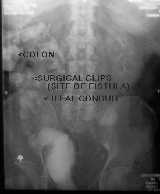
A subsequent IVP revealed a fistulous connection from high in the
ilieal conduit to the right colon at the site of surgical clips in
the right lower quadrant, suggesting breakdown of a prior anastomosis.
As urine leakage occurred late in the course of the IVP, it was thought
that urine diversion to the colon occurred only at higher pressures in
the neobladder/ileal conduit. Therefore, an indwelling catheter
was placed to keep the GU system decompressed, in hopes the fistula
would heal without further surgical intervention.
View followup image(gu).
Anterior delayed image from IVP
Differential Diagnosis List
Previous nuclear medicine study with tracer normally excreted via bowel.
Urine ingestion (Yes, its been reported!)
ACR Codes and Keywords:
References and General Discussion of Bone Scintigraphy (Anatomic field:Genitourinary System, Category:Neoplasm, Neoplastic-like condition)
Search for similar cases.
Edit this case
Add comments about this case
Return to the Teaching File home page.
Case number: bs089
Copyright by Wash U MO