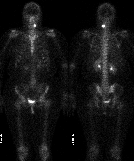

Anterior and posterior views
View main image(bs) in a separate image viewer
View second image(bs). Lateral view
View third image(ct). Computed tomography
Full history/Diagnosis is available below
Intense focus of activity left mid c-spine posteriorly. Otherwise normal.
2. Neck CT (9/10/97):
Expansile bony lesion of left pedicle of C4 with adjacent soft tissue changes.
Although osteoarthritis of an apophyseal joint is a possibility in this case based on bone scintigraphy alone, the patient’s young age, the degree of intense uptake and the single focus make this unlikely. The CT appearance excludes this.
Metastatic disease is a possibility. However, the patient has no history of cancer and this, in addition to the single focus of abnormal uptake, makes metastatic spread of tumor unlikely.
Osteochondroma (exostosis) may show normal to dramatically increased bone uptake. Osteochondroma is relatively uncommon in the spine (1-4% occur in the spine and account for 4% of solitary primary spine bone tumors). Thin-slice CT may provide the diagnosis if marrow and cortical continuity can be demonstrated. MRI will show the characteristic cartilagenous cap (intermediate on T1- and high on T2-weighted images). If this cap is greater than 1 to 2 cm in thickness, concern must be raised of chondrosarcoma. Normal to mild uptake in an exostosis generally excludes malignancy. However, marked uptake is not specific for malignant degeneration. More intense uptake is seen in growing children. Fracture, of course, results in intense uptake. Thus, the role of scintigraphy in exostosis or multiple hereditary exostosis is questionable.
Chondroblastoma is an uncommon benign cartilaginous bone lesion. It can result in normal to intense uptake. Ninety-percent occur between ages 5 and 25. It uncommonly involves the spine. These are usually eccentrically located in the epiphysis or apophysis, producing an osteolytic lesion that, in 30 to 50% of cases, will contain calcified foci.
Chondrosarcoma, the second most common malignant spinal primary bone tumor, is a destructive, lytic tumor with a chondroid matrix consisting of “rings and arcs” radiographically. Cortical destruction is always present (excluded by CT in this case). A soft tissue component is common. Scintigraphically, these produce patchy or homogeneous increased uptake.
Osteosarcoma is rare in the spine. The age group is older than that seen with osteosarcoma of the appendicular skeleton (typically fourth decade in spinal osteosarcoma). Scintigraphically, there is typically intense, patchy uptake.
Ewings sarcoma and PNET tumors are the most common primary spine malignant bone tumors in children. Most cases of spinal involvement are located in the sacrococcygeal region (they are relatively uncommon in the remainder of the spine). Radiographically these may produce permeative destruction, expansion or sclerosis. Scintigraphically, the tumor produces uptake in a pattern similar to that with osteomyelitis, with homogeneous uptake on immediate static and delay images. This is not the appearance seen in this case.
Other primary bone lesions and their scintigraphic appearance are described below:
Giant cell tumor of the spine is seen most commonly by a large margin in the sacrum, followed by thoracic, cervical and lumbar spine. Most lesions are benign. These are expansile, lytic, lesions which, in the spine, tend to involve the body of the vertebra. Scintigraphically, giant cell tumors commonly manifest as cold lesions with increased uptake around the rim (another cause of the “donut sign”). They may also have diffuse increased uptake.
Chordoma is the most common primary bone malignant tumor, other than lymhoproliferative tumors. They arise from notochord remnants, most commonly occurring near the midline in the region of the spheno-occipital synchondrosis or the sacrococcygeal region (but can occur anywhere in the spine). They are slow-growing, frequently large at diagnosis, and most common in the 30-60 year-old age group. Radiographically they are large, lytic lesions with a large soft-tissue component. The tumors may contain calcification.
Lymphoma and leukemia not uncommonly involve bone, either as a primary tumor, or more commonly as metastatic disease. Scintigraphy is not reliable in detecting these lesions. Most commonly they reveal increased uptake, but they may also be photopenic.
Enchondromas may have normal to mildly increased uptake. Marked uptake suggests malignant degeneration. They most commonly occur in the hands (usually proximal phalanges). It is rare in the spine. They are usually asymptomatic or may produce painless swelling. Pain should be considered a suspicious sign of malignant transformation.
Enostosis (bone island), although quite common in the spine, usually produces no or little increased uptake (enostoses larger than approximately 3 cm are more likely to be seen). These are more commonly seen in the body of a vertebra than posterior elements. Radiographically, these appear as a small sclerotic focus in the medullary cavity with spiculated margins. The expansile lesion seen on CT is not consistent with enostosis.
Fibrous cortical defects, such as non-ossifying fibroma, may have slight increased uptake. These have not been reported in the spine.
Eosinophilic granuloma may produce a range of increased uptake, usually mild, although large, aggressive lesions may show lack of uptake. Eosinophilic granuloma most commonly involves marrow of the skull and pelvic flat bones. It also commonly involves ribs and femora. In a vertebral body, it may produce vertebra plana. Thirty to forty percent of cases will produce no manifestations on bone scan. Bone scintigraphy and plain film skeletal survey are complementary exams in detection of bone involvement by EG.
Fibrous dysplasia will have moderately to markedly increased uptake, but will involve a more diffuse region of bone. Monostotic fibrous dysplasia rarely involves the spine (ribs are most common, followed by femur, tibia, mandible, calvarium and humerus). In the spine, it usually involves the body of the vertebra but can involve posterior elements.
Early bone infarct will produce a cold defect. During the reparative phase, it will become hot. The spine is an unusual location for this.
For table of bone lesions, see teaching points section below.
References:
Collier, B. David, et al. Skeletal Nuclear Medicine. Mosby year book. 1996.
Datz, Frederick, et al. Nuclear Medicine, A Teaching File. Mosby year book. 1992.
Frassica, Frank J., et al. Clinicopathologic features and treat
Follow-up CT reveals:
Interval osteoectomy of C4 inferior facet and bilateral C4 laminectomy.
Posterior spinal fusion C3-C7.
View followup image(ct). Follow-up CT
Marked uptake:
Moderate uptake:
Mild uptake:
Variable uptake:
Decreased uptake:
Bone destruction:
References and General Discussion of Bone Scintigraphy (Anatomic field:Skeletal System, Category:Neoplasm, Neoplastic-like condition)
Return to the Teaching File home page.