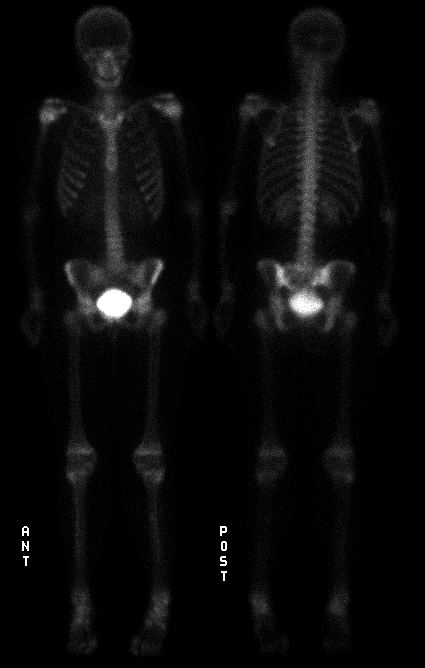

Anterior and posterior delayed whole body bone images
View main image(bs) in a separate image viewer
View second image(bs). Additional spot images (post-void images of the pelvis)
View third image(ct). Once you have decided on the region of bony abnormality, view this CT image for correlation.
Full history/Diagnosis is available below
The CT scan shows an expansile 6-cm soft tissue mass arising from the left sacrum. The mass obliterates the left sacral neural foramina and appears to break through sacral cortex, extending into the posterior pelvis.
"Cold defects" are often due to lytic tumors that do not incite significant reactive osteoblastic activity or to tumors (or other processes) that completely replace the bone marrow (and thus interfere with the blood supply to the bone). This appearance, though relatively uncommon, has been described in a wide variety of primary bone tumors and in metastases. At least 2% of metastatic lesions demonstrate decreased activity on bone scintigraphy and, although this finding is particularly common in multiple myeloma and renal cell carcinoma, one will encounter this appearance more frequently in association with breast and lung carcinoma given their overall greater incidence. Lesions of multiple myeloma, given their small size, may not be apparent on bone scintigraphy when they have decreased uptake.
This case is somewhat unusual due to the relatively young age of the patient, although GCT is certainly a consideration given the appearance and location of this tumor.
Reference: Skeletal Nuclear Medicine, B. David Collier, Ignac Fogelman, and Leonard Rosenthall, 1996
In retrospect, the original bone scintigraphy study performed 7 months previously was subtly positive (please see follow up image). There is a small region of decreased uptake in the left sacrum. The interval change since the prior exam highlights the degree of bony destruction in the sacrum.
View followup image(bs). Posterior view of the pelvis (original bone scintigraphy)
Sacral osteomyelitis with bony destruction (in the setting of a decubitus ulcer)
Osteoradionecrosis (following radiation therapy)
References and General Discussion of Bone Scintigraphy (Anatomic field:Spine and Contents, Category:Neoplasm, Neoplastic-like condition)
Return to the Teaching File home page.