Case Author(s): David Hillier, M.D., Ph.D. and Tom R. Miller, M.D., Ph.D. , 10/7/97 . Rating: #D2, #Q3
Diagnosis: Reparative bone / Intra-operative probe with autoradiograph of surgical specimen
Brief history:
This is a 49 year-old woman with a history of breast cancer,
Images:

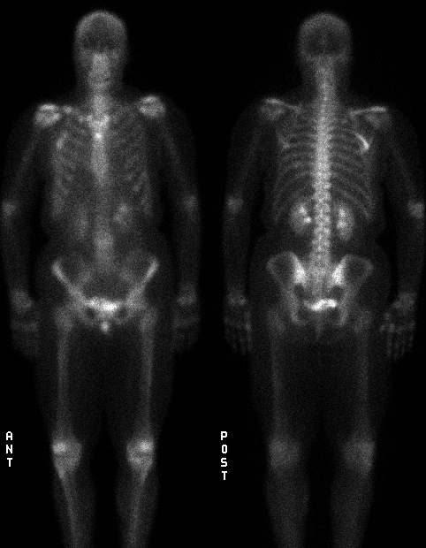
Anterior and posterior views
View main image(bs) in a separate image viewer
View second image(bs).
Right anterior oblique view
View third image(bs).
Bone scintigraphy of surgical specimen
View fourth image(mr).
MRI of right fifth rib
Full history/Diagnosis is available below
Diagnosis: Reparative bone / Intra-operative probe with autoradiograph of surgical specimen
Full history:
This is a 49 year-old woman with a history of breast cancer diagnosed two months ago and treated by right mastectomy one month ago. The surgical
margins were positive. Bone scintigraphy was performed. She
subsequently underwent chemotherapy and then stem cell bone marrow
transplant. She also has a history of a motor vehicle accident 30 years
ago with right clavicle fracture, multiple rib fractures, punctured lung
and splenic laceration.
Radiopharmaceutical:
20.2 mCi Tc-99m methylene diphosphonate
Findings:
1. Bone scintigraphy (7/8/97) 1 month after right mastectomy:
- Long segment of uptake in a posterolateral right rib with no
radiographic correlate. There is a focus of mildly increased uptake
corresponding to an old right mid-clavicle fracture.
(2). Right rib films (7/8/97) a(not included):
- Revealed no rib abnormality. There was an old healed right
clavicle fracture.
3. MRI of chest (7/18/97):
- Reveals abnormal signal (low on T1 and high on T2) in the marrow of
the right posterolateral fifth rib. The differential diagnosis includes fibrous
dysplasia, but this is virtually excluded due to the normal plain film.
This was felt to most likely represent metastatic disease.
4. Bone scintigraphy and rib surgical specimen autoradiograph (7/23/97):
- Tc-99m MDP was injected prior to surgical removal of the right
5th rib. Intraoperative gamma probe localization was used. The
resected surgical specimen was then brought to the nuclear medicine
department where a scintigraphic image was obtained. This revealed
increased uptake in the anterior 2/3 of the specimen w.r.t. the
posterior 1/3. A single image of the right hemithorax reveals that a
portion of the abnormal 5th rib has been resected but that a portion of
the abnormal region of uptake remains at the lateral aspect.
(5). CT of chest, abdomen and pelvis (11/14/97) (not included):
- Three new indeterminant pulmonary nodules worrisome for metastases.
- Bilateral adrenal masses. Stable metastases cannot be excluded.
Discussion:
The technique of using a radioactive tracer as a label and then
obtaining an image of the spatial distribution of radioactivity from the
specimen with radiographic film is known as autoradiography. In this
case, the surgical specimen was obtained after administration of a
bone-seeking radiopharmaceutical and the image was obtained
scintigraphically.
The technique of administering a bone radiopharmaceutical prior to
surgery for removal of an osteoid osteoma has been described. The
surgeon can use a gamma probe to assist in intra-operative localization
of the nidus and aid conservative excision. The pathologist can use
autoradiography to help identify small lesions. Bone chips are placed
over x-ray film with an intensifying screen for 2 - 8 hours.
This case illustrates the use of autoradiography, obtained
scintigraphically, to verify that the resected specimen contains the
region of interest. This is an unusual application for bone
scintigraphy since it is also logistically difficult (the surgery must
follow the bone scintigraphy injection within approximately 24 hours, and yet
the need for surgery may not be known until the examination is finished).
Nonetheless, this technique can prove helpful given the right
circumstances.
References:
Ghelman, Bernard and Vigorita, Vincent. Postoperative Radionuclide
Evaluation of Osteoid Osteomas. Radiology: 146; 509-512, 1983.
Vigorita, VJ and Ghelman, B. Localization of Osteoid Osteomas - Use
of Radionuclide scanniing and autoimaging in identifying the nidus. Am
J Clin Path. 79: 2; pp 223-226, 1983.
Followup:
Pathology on the rib specimen reveals no evidence of cancer.
There was a large area of reparative bone. Remodeling was present,
characterized by focal osteoclastic activity. The new bone did not
reveal active osteoblastic activity. These findings are therefore most
consistent with a trauma-induced lesion.
Seven months after these studies were performed the patient
underwent stem cell bone marrow transplant.
Differential Diagnosis List
The differential diagnosis of increased uptake in a long segment
of rib includes metastatic disease, fibrous dysplasia (largely exlcuded
by the normal appearance on plain film), osteonecrosis (there was no
radiation therapy in this case and the posterior location is atypical),
osteomyelitis (the clinical history does not suggest this) or an
adjacent soft tissue inflammatory process (also not supported by
history). The patient has a distant history of trauma. Usually, trauma
manifests as a focal area of increased uptake. However, in this case,
the patient had sustained severe trauma with multiple rib fractures,
punctured lung, splenic laceration and clavicle fracture. Thus,
reparative bone over a longer segment of bone than usual is a
consideration. There is a mild focal uptake in the right mid clavicle
corresponding in location to a prior fracture 30 years ago (this may be
due to reparative bone, or to altered morphology of the healed bone with
increased thickness at the site).
ACR Codes and Keywords:
References and General Discussion of Bone Scintigraphy (Anatomic field:Skeletal System, Category:Neoplasm, Neoplastic-like condition)
Search for similar cases.
Edit this case
Add comments about this case
Return to the Teaching File home page.
Case number: bs083
Copyright by Wash U MO

