Case Author(s): Stephanie P.F. Yen, M.D. and Keith Fischer, M.D. , 09/26/97 . Rating: #D2, #Q4
Diagnosis: Polyostotic fibrous dysplasia
Brief history:
Eight-year-old female with right hip pain.
Images:
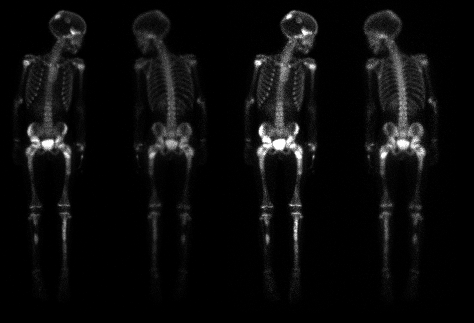
Anterior and posterior whole body scintigrams are shown.
View main image(bs) in a separate image viewer
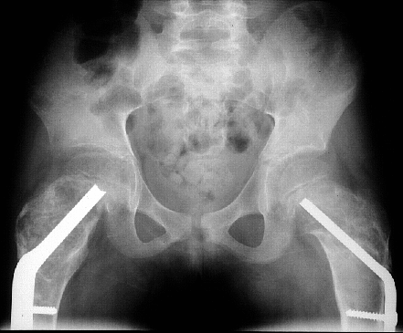
View second image(xr).
Anteroposterior view of the pelvis.
Full history/Diagnosis is available below
Diagnosis: Polyostotic fibrous dysplasia
Full history:
Eight-year-old female with polyostotic fibrous dysplasia who is status
post bilateral valgus osteotomies of the hips in 12/96. She presents
with a two week history of right hip pain and limp.
Radiopharmaceutical:
6.0 mCi Tc-99m MDP, intravenously
Findings:
The anterior and posterior whole-body scintigrams demonstrate multiple
bilateral areas of abnormal radiopharmaceutical uptake in the axial and appendicularappendicular
skeleton. Specifically, these areas include the skull and both iliac
wings, proximal and mid femurs, and tibias, which demonstrate fairly
symmetrical uptake. There is asymmetric increased activity involving the
acetabular regions, right greater than left. No definite abnormal
activity is appreciated in either femoral head to suggest avascular necrosis.
Linear areas of decreased activity are seen in both proximal femurs and
traversing both femoral necks, and are compatible with hardware from the
patient's known bilateral hip osteotomies.
The anteroposterior radiograph of the pelvis demonstrates bubbly-appearing
cystic lesions involving both ilia, right greater than left, and both
proximal femurs. In addition, note is made of expansion of the right
supra-acetabular area. The radiographic appearance is consistent with the
patient's history of polyostotic fibrous dysplasia, and the areas of
radiographic abnormality correlate with those areas of abnormal
radiopharmaceutical uptake seen in the pelvis on the bone scintigraphy
study. The patient is noted to be status post bilateral valgus hip
osteotomies.
Discussion:
The scintigraphic features of fibrous dysplasia, a benign developmental disorder of bone-forming mesenchyme, are nonspecific. Fibrous dysplasia will demonstrate hyperemia, increased blood pool activity, and relatively intense radiopharmaceutical uptake on the delayed images. However, the diagnosis can often be made with correlation with radiographs, where the lesions classically demonstrate radiolucency with ground glass appearance, although the radiographic appearance of fibrous dysplasia can be quite variable.
The majority of cases, approximately 70-80%, are monostotic, while the remaining 20-30% are polyostotic. 2-3% of patients with fibrous dysplasia have associated endocrinopathies. The ribs, femur, tibia, calvarium, bones, spine, pelvis, and shoulder girdle are frequently involved. Polyostotic fibrous dysplasia predominantly is unilateral in its distribution, however, extensive polyostotic fibrous dysplasia, as demonstrated in this case, is commonly bilateral, albeit asymmetric, in distribution.
References: 1. Feldman F. Tuberous sclerosis, neurofibromatosis, and fibrous dysplasia. In: Resnick D and Niwayama G, ed. Diagnosis of Bone and Joint Disorders, 2nd ed. WB Saunders Company, Philadelphia, PA, 1988: 4057-4070. 2. Mandell GA and Harcke HT. Pediatric bone scanning. In: Collier BD, Fogelman I, and Rosenthall L, ed. Skeletal Nuclear Medicine. Mosby, St. Louis, MO, 1996: 343-344.
Major teaching point(s):
Bone scintigraphy is useful in establishing the diagnosis of polyostotic fibrous dysplasia.
ACR Codes and Keywords:
- General ACR code: 48
- Skeletal System:
4.85 "FIBROUS DYSPLASIA"
References and General Discussion of Bone Scintigraphy (Anatomic field:Skeletal System, Category:Misc)
Search for similar cases.
Edit this case
Add comments about this case
Read comments about this case
Return to the Teaching File home page.
Case number: bs082
Copyright by Wash U MO