
After viewing the image(s), the Full history/Diagnosis is available by using the link here or at the bottom of this page
Anterior and posterior whole body scintigrams are shown.
View main image(bs) in a separate viewing box
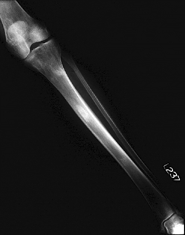
View second image(xr). Anteroposterior view of the left tibia and fibula.
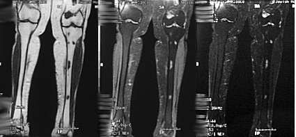
View third image(mr). Coronal T1, proton density, and T2-weighted images of the bilateral lower extremities.
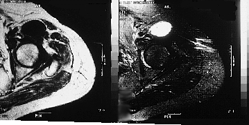
View fourth image(mr). Axial T1 and T2-weighted FSE images of the left hip.
Full history/Diagnosis is also available
Return to the Teaching File home page.