Case Author(s): Lisa Oakley M.D. and Tom R. Miller M.D.,Ph.D. , 07/11/97 . Rating: #D3, #Q3
Diagnosis: Soft tissue hematoma
Brief history:
Elderly woman with hip pain after a fall.
Images:
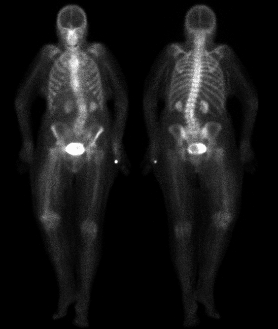
ANTERIOR AND POSTERIOR DELAYED WHOLE BODY IMAGES
View main image(bs) in a separate image viewer
Full history/Diagnosis is available below
Diagnosis: Soft tissue hematoma
Full history:
94 year-old female who fell off the toilet this morning. The patient has
a persistent externally rotated right leg and refuses to move.
Radiographs of the right femur are reportedly negative from an outside
hospital.
Radiopharmaceutical:
Tc-99m MDP
Findings:
There is faint, focally increased radiopharmaceutical uptake in the region
of the right femoral greater trochanter. There is also diffusely
increased soft tissue uptake in the right thigh. The right thigh
appears asymmetrically enlarged when compared with the
left thigh.
Discussion:
Skeletal fractures incite bone repair mechanisms usually
within 24 hours of injury. This change (initially due to hyperemia)
is seen on bone scans as focal increased activity. Matin found that
80% of patients with fractures have a positive bone scan by 24 hours
and 92% by 72 hours. However, in elderly patients bone
scans may take longer to become postive and also may return to normal more slowly.
Therefore, in older patients with suspected fracture and negative initial
studies (e.g. plain film, MRI, bone scan),
repeat bone scan at 72 hours may unveil lesions which were previously
occult.
Followup:
The patient continued to have pain, and an MRI was ordered the following
day.
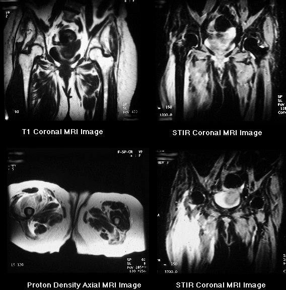
MRI images of the hips demonstrate normal bone marrow signal and no
evidence of acute fracture. However, there is extensive right thigh
soft tissue swelling and hematoma which is most impressive around the
anterior and medial muscle compartments. This is seen as areas of
increased signal on the proton density and STIR sequences, and
intermediate signal on T1-weighted images.
View followup image(mr).
MRI IMAGES OF THE HIPS
Major teaching point(s):
This study may have been falsely negative for fracture.
Repeat bone scintigraphy at 72 hours may have been helpful because
of the relatively long interval between fracture and the appearance of
scintigraphic abnormalities, especially in elderly patients. One school
of thought recommends deferring initial bone scintigraphy until at least 24
hours after the suspected fracture in older patients. Others recommend
proceeding with imaging because the study might be positive or, as in this
unusual case, reveal other abnormalities. In any case, bone scintigraphy
could be repeated at a later date if initially negative.
The study was valuable, nevertheless, because the soft tissue findings
suggested post-traumatic hematoma later confirmed by MRI.
ACR Codes and Keywords:
- General ACR code: 44
- Skeletal System:
4.49 "MISCELLANEOUS"
References and General Discussion of Bone Scintigraphy (Anatomic field:Skeletal System, Category:Effect of Trauma)
Search for similar cases.
Edit this case
Add comments about this case
Read comments about this case
Return to the Teaching File home page.
Case number: bs078
Copyright by Wash U MO