Case Author(s): Anton J. Johnson, M.D., Ph.D. and Jerold W. Wallis, M.D. , 5/1/97 . Rating: #D2, #Q4
Diagnosis: Liver metastases
Brief history:
Elderly female with back pain.
Images:
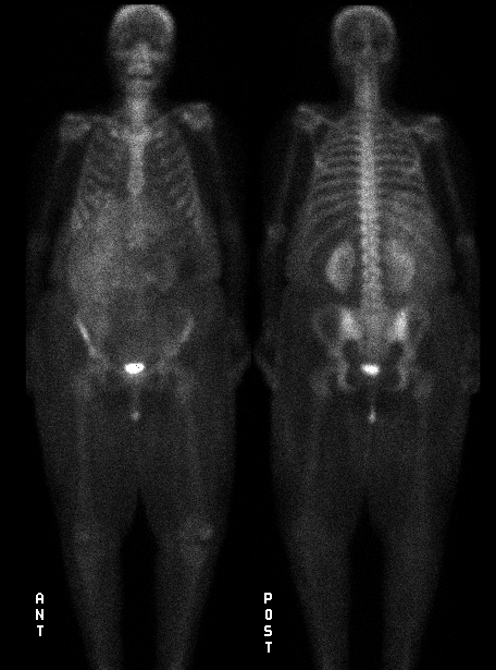
Anterior and posterior whole body images.
View main image(bs) in a separate image viewer
Full history/Diagnosis is available below
Diagnosis: Liver metastases
Full history:
Elderly female with history of colon carcinoma, status post
colectomy in the past.
Radiopharmaceutical:
Tc-99m MDP
Findings:
Soft tissue uptake is seen throughout an enlarged liver. There
is no evidence of metastatic disease to bone.
Discussion:
Diffuse abnormal
liver uptake can be due to a radiopharmaceutical problem (with colloid
formation), severe liver ischemia with patchy infarction (similar
to pyrophosphate uptake in the heart during myocardial infarct
imaging), and associated with infarction/microcalcification
due to extensive liver metastatic disease.
Followup:
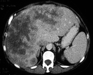
Computed tomography examination demonstrated extensive liver
metastases.
View followup image(ct).
Image from a contrast enhanced CT of abdomen.
ACR Codes and Keywords:
References and General Discussion of Bone Scintigraphy (Anatomic field:Skeletal System, Category:Neoplasm, Neoplastic-like condition)
Search for similar cases.
Edit this case
Add comments about this case
Return to the Teaching File home page.
Case number: bs074
Copyright by Wash U MO