Case Author(s): Scott Winner, M.D. and Jerold Wallis, M.D. , 3-21-97 . Rating: #D2, #Q5
Diagnosis: osteomtelitis of the proximal phalynx of the right second toe.
Brief history:
69-year old woman with pain in
right second toe.
Images:
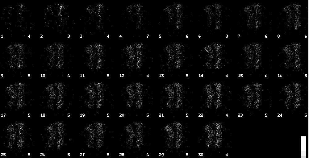
Plantar flow image of feet
View main image(bs) in a separate image viewer
View second image(bs).
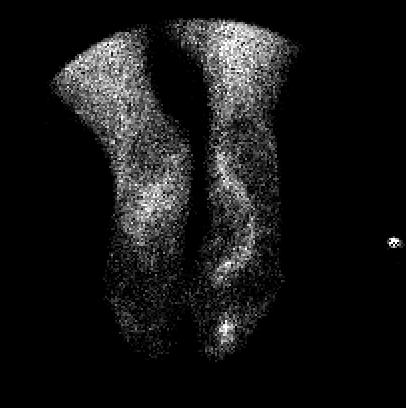
Immediate static images
View third image(bs).
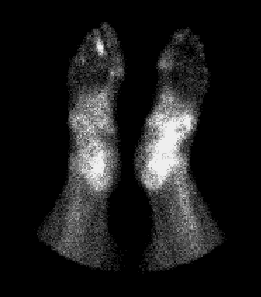
Two hour delay static images
View fourth image(xr).
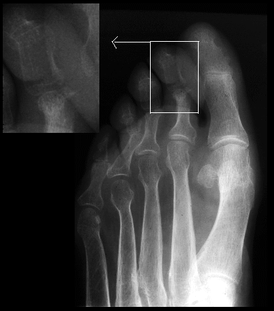
Radiograph of right foot
Full history/Diagnosis is available below
Diagnosis: osteomtelitis of the proximal phalynx of the right second toe.
Full history:
69-year old woman who removed
a corn from her right second toe in November, 1996.
She presents with chronic purulent drainage.
Radiopharmaceutical:
21.0 mCi Tc-99m MDP
i.v.
Findings:
There is moderately to markedly
increased radiopharmaceutical activity noted on the
angiographic, blood pool, and delayed static images in
the distal aspect of the proximal second phalanx of the
right foot. This is most consistent with osteomyelitis.
Discussion:
Three-phase imaging is a
technique used to help differentiate cellulitis from
osteomyelitis. The distinction is clinically important
because of the therapeutic implications of prolonged
treatment when osteomyelitis is diagnosed. Cellulitis
demonstrates hyperemia with increased activity
diffusely on the blood pool images and subsequent
clearance of tracer on the delayed images without
focally increased uptake in bone. Typically,
osteomyelitis shows increased blood flow with
focally or diffusely increased uptake of tracer on the
blood pool images. Osteomyelitis differs from
cellulitis on the delayed images in that progressive
focal accumulation of tracer is seen within the
involved bone when osteomyelitis is present. This
case demonstrates findings classic for osteomyelitis.
A correlative radiograph of the right foot was obtained
after bone scintigraphy was performed. This
radiograph demonstrated bony destruction in the
medial aspect of the second proximal phalanx
corresponding in location to the abnormal
accumulation of tracer seen on the blood pool and
delayed images.
Differential Diagnosis List
Increased uptake on all three phases of the bone scintigraphy
can be seen in other disorders, including neuropathic
joint disease, gout, fracture, severe arthritis, healing
osteonecrosis, and bone tumors. Therefore, it is
imperative that correlation with clinical data and
other imaging studies be performed in conjunction
with the three-phase bone scintigraphy.
ACR Codes and Keywords:
- General ACR code: 42
- Skeletal System:
4.21 "OSTEOMYELITIS (for 4th number see box following .219)"
References and General Discussion of Bone Scintigraphy (Anatomic field:Skeletal System, Category:Inflammation,Infection)
Search for similar cases.
Edit this case
Add comments about this case
Read comments about this case
Return to the Teaching File home page.
Case number: bs073
Copyright by Wash U MO