Case Author(s): Anton J. Johnson, M.D., Ph.D. and Keith Fischer, M.D. , 11/8/96 . Rating: #D2, #Q5
Diagnosis: Camera off peak (Co-57 instead of Tc-99m)
Brief history:
81 year old woman with right hip pain after a recent fall.
Images:

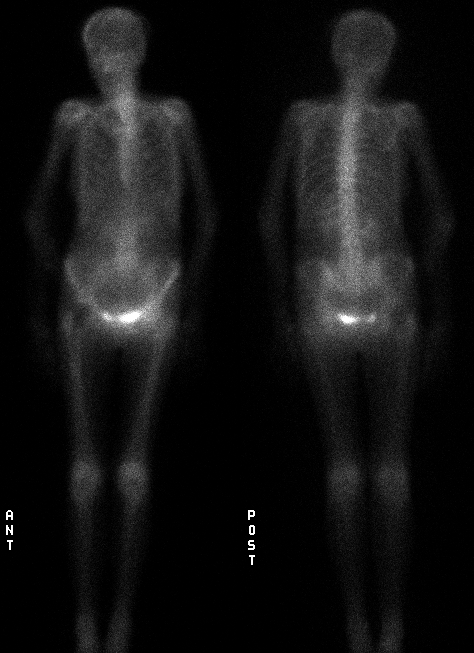
Delayed anterior and posterior whole body images.
View main image(bs) in a separate image viewer
Full history/Diagnosis is available below
Diagnosis: Camera off peak (Co-57 instead of Tc-99m)
Full history:
81 year old woman with right hip pain after a recent fall.
Radiopharmaceutical:
20.4 mCi Tc-99m MDP i.v.
Findings:
Apparent diffusely increased soft tissue activity with poor
visualization of the bones. This appearance raised the question
of a technical problem. The gamma camera was checked and its energy
peak found to be set erroneously low at 122 keV, the energy of
Co-57.
Discussion:
Co-57 has a relatively long half-life (270 days) and an energy peak
(122 keV) similar to that of Tc-99m, two attributes that make it
desirable for use in quality control. At our institution, Co-57 flood
sources are used for daily field homogeneity quality control. A minor
drawback is that you have to remember to reset the energy peak of the
gamma camera before you begin imaging patients. If this is not done,
subsequent studies using Tc-99m (or higher energy) labeled compounds
will yield low resolution images with apparent poor radiopharmaceutical
localization. This is because the incorrectly set camera is imaging
lower energy photons that have been scattered (Compton) from random
sites throughout the patient's body. Normally, most of these lower
energy Compton scattered photons are rejected (not used for image
construction) when the camera's energy peak and window are set
properly.
An alternative to Co-57 is to use a solution containing Tc-99m
pertechnetate as the flood source. Although this obviates the need to
reset the energy peak for subsequent Tc-99m studies, a new solution has
to be prepared every day because of Tc-99m's six hour half-life. The
added work of daily preparation, along with the potential problems
which can arise if the Tc-99m pertechnetate solution is not homogeneous
or of uniform thickness, have led many nuclear medicine departments to
prefer Co-57 flood sources.
Followup:
The camera's energy peak was reset to the correct value used for Tc-99m
containing radiopharmaceuticals (140 keV) and the patient re-imaged.
The findings include a right femoral prosthesis with no evidence of
fracture.
View followup image(bs).
Delayed anterior and posterior images with the camera
set on the correct peak (140 keV).
Major teaching point(s):
Do not forget to include 'camera off-peak' in the differential
diagnosis of a bone scan with apparent poor skeletal radiopharmaceutical
uptake.
Differential Diagnosis List
Once incorrect energy peak has been excluded, the general differential
diagnosis for poor skeletal localization of Tc-99m MDP includes poor
radiopharmaceutical preparation, congestive heart failure,
bisphosphonate therapy, osteoporosis, and incorrect radiopharmaceutical
administration (e.g., Tc-99m DTPA instead of Tc-99m MDP for bone
scintigraphy).
ACR Codes and Keywords:
References and General Discussion of Bone Scintigraphy (Anatomic field:Skeletal System, Category:Normal, Technique, Congenital Anomaly)
Search for similar cases.
Edit this case
Add comments about this case
Read comments about this case
Return to the Teaching File home page.
Case number: bs069
Copyright by Wash U MO

