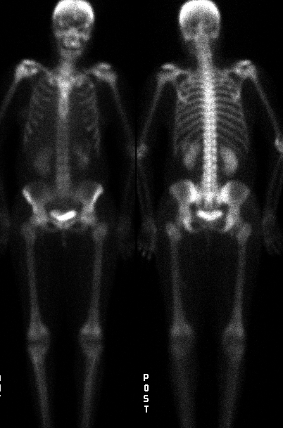

Anterior and posterior whole-body delayed images.
View main image(bs) in a separate image viewer
View second image(bs). Lateral view of the skull.
View third image(bs). Oblique view of the chest including the right arm.
Full history/Diagnosis is available below
Complaints of bone and joint pain are common in patients with several types of sickle cell hemoglobinopathies. Symptomatic episodes are usually transient, occur during crisis, and resolve without producing radiologic change. The symptoms are often due to bone marrow infarction with or without secondary involvement of the bony structures. Radionuclide scintigrams are sensitive for early detection. They also aid in the evaluation of the extent of damage to the bone and marrow.
Some investigators feel that the initial evaulation of sickle cell patients presenting with bone pain should be done with bone marrow scintigraphy, as it may be more useful than bone scintigraphy for differentiating infarct from osteomyelitis in these patients.
Uptake of the bone scan agent in the spleen in sickle cell disease is quite common by the end of the first decade. Uptake takes place in areas of infarction with revascularization, but is not uniformly seen in all patients. Repetitive infarcts lead to deposition of calcium salts and iron which, in turn, cause uptake the bone agent. Uptake of these agents by the spleen in sickle cell disease correlates with the presence of functional asplenia.
REFERENCE: Nuclear Medicine. Henkin et al., editor. Mosby, 1996.
View followup image(ct). Small, calcified spleen in the left upper quadrant.
References and General Discussion of Bone Scintigraphy (Anatomic field:Skeletal System, Category:Other generalized systemic disorder)
Return to the Teaching File home page.