Case Author(s): Samuel Wang, M.D. and Jerold Wallis, M.D. , 9/22/96 . Rating: #D3, #Q3
Diagnosis: Spinous process contusions/fractures.
Brief history:
40-year old woman who
fell from a horse approximately seven weeks
ago and now has complaints of back pain.
Images:

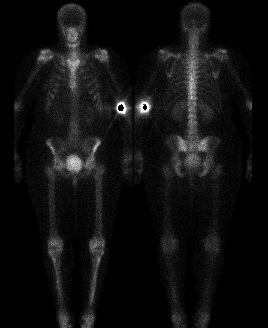
Anterior and posterior whole body images.
View main image(bs) in a separate image viewer
View second image(bs).
SPECT sagittal images.
Full history/Diagnosis is available below
Diagnosis: Spinous process contusions/fractures.
Full history:
This 40-year old woman
fell from a horse approximately seven weeks
prior to this examination. She landed "flat on
her back" and has had persistent complaints of
thoracolumbar back pain.
Radiopharmaceutical:
Tc-99m MDP
Findings:
The whole-body planar images
demonstrate mildly increased
radiopharmaceutical uptake in the spinous
processes of T7-11. This is seen much more
clearly on the sagittal SPECT reconstructed
images. Additionally, there is moderately
increased uptake in the T7 vertebral body. The
spinous processes were not well visualized on
plain films although mild anterior compression
of the vertebral body of T7 was noted.
Discussion:
Given the mechanism of
injury and the appearance of consecutive foci
of uptake, these findings are most consistent
with contusion and/or fractures of the spinous
processes of T7-11 as well as compression
fracture of T7.
Increased uptake in the posterior spinous processes was quite subtle on the
original film version of the planar images, though it can be seen on careful
examination of the digital images shown here. Oblique planar images or
SPECT both can improve visualization of this site.
ACR Codes and Keywords:
References and General Discussion of Bone Scintigraphy (Anatomic field:Skeletal System, Category:Effect of Trauma)
Search for similar cases.
Edit this case
Add comments about this case
Read comments about this case
Return to the Teaching File home page.
Case number: bs064
Copyright by Wash U MO

