Case Author(s): Anton J. Johnson, M.D., Ph.D. and Tom R. Miller, M.D., Ph.D. , 7/26/96 . Rating: #D2, #Q3
Diagnosis: Metabolic bone disease secondary to renal failure.
Brief history:
17 year-old male with fatigue, weight loss, and fever.
Images:

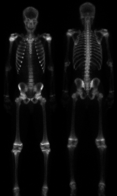
Anterior and posterior images are shown.
View main image(bs) in a separate image viewer
Full history/Diagnosis is available below
Diagnosis: Metabolic bone disease secondary to renal failure.
Full history:
17 year-old male patient with
fatigue, weight loss, and fever. Laboratory results
include elevated BUN, creatinine, and phosphate
with decreased serum levels of calcium. The patient
also has a history of chronic renal disease.
Radiopharmaceutical:
18.2 mCi Tc-99m
MDP i.v.
Findings:
Whole-body bone scintigraphy
demonstrates diffusely increased skeletal uptake, minimal soft tissue and renal uptake and no bladder activity.
There are no focal bony lesions.
Discussion:
The most important aspect
of interpretation of a "superscan" on
bone scintigraphy is to properly identify it as
abnormal -- the lack of focal bony abnormalities
tends to be misleading. Near total absence of renal
and soft tissue activity, however, should prompt the
interpreter to consider the presence of a superscan.
The differential diagnosis for a true superscan is
essentially limited to metabolic versus diffuse
metastatic disease. The word "true" is included
because a superscan appearance can be artificially
obtained by a prolonged delay before imaging (e.g.
6 or more hours rather than the usual 2-3 hours after
injection). The additional time allows for more
renal and soft tissue clearance of tracer with
resultant greater bone-to-soft tissue activity.
Fortunately, this is an infrequent problem and one
that can be identified by noting an abnormally long
delay from time of injection to imaging.
Given a true superscan, clinical history is
frequently all that is necessary to suggest the correct
diagnosis. Prostate carcinoma is the most common
of the metastatic causes, with breast and lymphoma
as other less likely possibilities. Renal disease and
hyperparathyroidism (primary or secondary) lead
the list of metabolic causes of superscans. Typical
histories would include an elderly male with a
significantly elevated PSA (suggesting metastatic
prostate carcinoma) or a patient with chronic renal
failure and/or laboratory evidence of renal disease
(suggesting a metabolic etiology). In this teaching
file case, the patient's young age, history of chronic
renal disease, and abnormal laboratory values
clearly point to metabolic disease as the cause of his
superscan. Without this clinical information,
however, metastatic disease would remain in the
differential, although widespread metastases
typically show less involvement of the extremities
and are usually not completely uniform and symmetric compared to metabolic
bone disease.
References: Datz FL: Handbook of Nuclear
Medicine, 2nd ed. St. Louis, Mosby, 1993,
pp67-69. Datz FL, et al: Nuclear Medicine, A
Teaching File, St. Louis, Mosby, 1992, p26. Mettler FA, Guiberteau MJ: Essentials
of Nuclear Medicine, 3rd ed. Philadelphia,
W.B. Saunders, 1991, p217. Thrall JH, Ziessman HA: Nuclear
Medicine, the Requisites, St. Louis, Mosby,
1995, pp102,120.
Major teaching point(s):
See Discussion
Differential Diagnosis List
See Discussion
ACR Codes and Keywords:
References and General Discussion of Bone Scintigraphy (Anatomic field:Skeletal System, Category:Metabolic, endocrine, toxic)
Search for similar cases.
Edit this case
Add comments about this case
Return to the Teaching File home page.
Case number: bs062
Copyright by Wash U MO

