Case Author(s): Jerold W. Wallis , 5/11/96 . Rating: #D1, #Q4
Diagnosis: Sacral fracture
Brief history:
Low back/pelvic pain
Images:

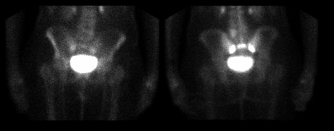
Anterior and posterior images of the pelvis
View main image(bs) in a separate image viewer
Full history/Diagnosis is available below
Diagnosis: Sacral fracture
Full history:
The patient is an elderly woman with no history of
malignancy, who has had low back/pelvic pain since
a fall two weeks ago. Radiographs of the pelvis,
hips, and lumbar spine demonstrate only degenerative
changes in the lumbar spine, and old lumbar compression
fractures.
Radiopharmaceutical:
20 mCi Tc-99m MDP, i.v.
Findings:
Markedly increased uptake is seen in the mid sacrum
in a transverse linear distribution, likely also
involving the inferior aspect of the sacro-illiac
joints.
Discussion:
The findings are diagnostic of a sacral fracture.
Major teaching point(s):
Bone scintigraphy may reveal a fracture which is not
evident on plain radiographs.
The appearance of sacral insufficiency fractures is
variable; sometimes the vertical components in the
sacroiliac joints are more pronounced, giving
an "H" configuration to the fracture zone.
ACR Codes and Keywords:
References and General Discussion of Bone Scintigraphy (Anatomic field:Skeletal System, Category:Effect of Trauma)
Search for similar cases.
Edit this case
Add comments about this case
Read comments about this case
Return to the Teaching File home page.
Case number: bs059
Copyright by Wash U MO

