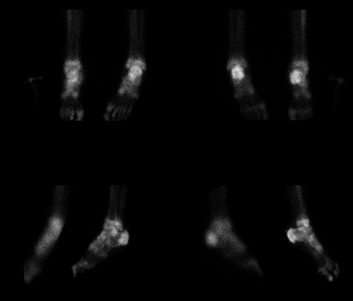
Anterior, posterior, and lateral delayed images of the feet.
View main image(bs) in a separate image viewer
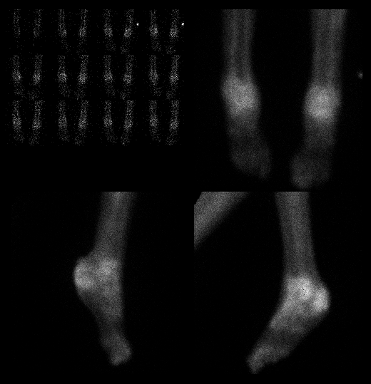
View second image(bs). Flow and immediate images of the feet. The flow images and upper right immediate image are posterior views.
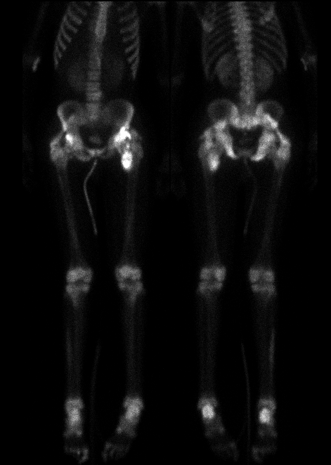
View third image(bs). Anterior and posterior images of the whole body. Can you figure out the patient's underlying medical problem?
Full history/Diagnosis is available below
Imaging of the whole body demonstrate increased activity around the hip joints in a distribution not corresponding to normal bone; this finding is consistent with myositis ossificans in the paraplegic patient.
Increased activity can be seen in the mid sacrum as well, with probable increased activity at the ischial tuberosities.
Other possible causes of increased uptake on a three-phase bone scan include fracture, healing avascular necrosis, bone involvement with primary or metastatic tumor, and Paget's disease. Mild diffuse increased uptake on delayed imaging can also be seen in the setting of increased blood flow from overlying cellulitis.
Note that evaluation of the sacrum can be difficult, since the normal appearance can vary due to the variable angulation of the sacrum. In this patient, the increased uptake in the mid sacrum and absent uptake in the lower sacrum warrented careful inspection of plain radiographs of this region (see below).
References and General Discussion of Bone Scintigraphy (Anatomic field:Skeletal System, Category:Inflammation,Infection)
Return to the Teaching File home page.