Case Author(s): Samuel Wang, M.D. , . Rating: #D3, #Q3
Diagnosis: Dermatomyositis
Brief history:
76-year old man with
complaints of neck pain and neck and bilateral upper
extremity weakness. The patient also has a skin rash.
Images:

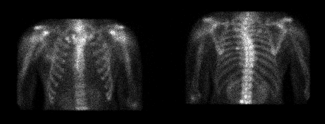
Anterior and posterior images.
View main image(bs) in a separate image viewer
View second image(xr).
Anterior chest.
View third image(ct).
CT of liver.
Full history/Diagnosis is available below
Diagnosis: Dermatomyositis
Full history:
76-year old man admitted to the
hospital with complaints of dysphagia and
gastrointestinal bleeding. The patient also has
complaints of neck pain as well as neck and bilateral
upper extremity weakness. The patient has been
unable to raise his hands over his head for the last
month. Upper endoscopy and biopsy demonstrated
gastric antral adenocarcinoma. The patient also had
prostate biopsy, which demonstrated adenocarcinoma.
Abnormal laboratory values included CK of 1653
(normal 30-22), LDH of 441 (normal 100-250), and
SGOT of 173 (normal 11-47). PSA was 93. This
patient also had a maculopapular, scaling, eczematous
skin rash.
Radiopharmaceutical:
Tc-99m MDP
Findings:
Bone scintigraphy demonstrates
diffuse soft tissue uptake around the neck and
shoulders. Plain film of the chest demonstrates no
calcifications in the soft tissue, although there is some
prominence of the soft tissues in the shoulder girdle.
The CT study demonstrates liver metastases.
Discussion:
Common and uncommon causes of
skeletal muscle uptake on bone scintigraphy are
electrical burns (including cardioversion), muscle
trauma, myositis ossificans, peripheral vascular
disease with ischemia, rhabdomyolysis,
polymyositis/dermatomyositis, iron dextran injection,
and calcific tendinitis.
This patient's shoulder pain and weakness is
consistent with polymyositis but is not specific. With
the characteristic skin rash, however,
dermatomyositis is most likely. The dermatomyositis
is almost certainly paraneoplastic in this patient with
gastric adenocarcinoma and hepatic metastases.
There are five types of poly/dermatomyositis. Type I
is primary idiopathic myositis. Type II is primary
idiopathic dermatomyositis. Type III is either,
associated with neoplasm (approximately 10% of
cases), especially in older patients. Type IV is either,
associated with vasculitis in children. Type V is
either, associated with collagen vascular disease
(approximately 30% of cases).
Followup:
The patient underwent partial
gastrectomy and will undergo chemotherapy, which
should also treat the dermatomyositis. No specific
treatment for the dermatomyositis is planned
(steroids are the usual form of treatment).
ACR Codes and Keywords:
References and General Discussion of Bone Scintigraphy (Anatomic field:Skeletal System, Category:Other generalized systemic disorder)
Search for similar cases.
Edit this case
Add comments about this case
Read comments about this case
Return to the Teaching File home page.
Case number: bs056
Copyright by Wash U MO

