Case Author(s): Samuel Wang, M.D. and Barry A. Siegel, M.D., . Rating: #D3, #Q3
Diagnosis: Poorly differentiated infiltrating ductal breast carcinoma with metaplastic changes.
Brief history:
61-year old woman with a large breast
mass
Images:

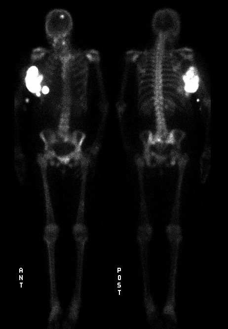
Anterior and posterior whole-body images.
View main image(bs) in a separate image viewer
View second image(bs).
Oblique and lateral chest images.
View third image(ct).
CT of the chest.
Full history/Diagnosis is available below
Diagnosis: Poorly differentiated infiltrating ductal breast carcinoma with metaplastic changes.
Full history:
61-year old woman with a history
of breast cancer diagnosed four years previously. The
patient refused surgery at that time and she presents
currently with a large fungating mass involving the
right breast. The tumor is adherent to the chest wall.
In addition, the patient was in a motor vehicle
accident two years previously at which time she
sustained multiple pelvic fractures.
Radiopharmaceutical:
Tc-99m MDP
Findings:
A large focal area of intensely
increased uptake is identified in the right breast.
This corresponds to the known fungating partially
calcified tumor mass. The tumor on bone scintigraphy
consists of a large mass with two small satellite
components seen anteriorly. Chest radiograph and
CT confirmed the presence of calcification within the
large breast mass.
Areas of increased activity also are identified in the
left parietal bone, the left scapula, and the mid shafts
of both femora, consistent with metastatic lesions.
Several areas of increased uptake also are identified
in the pelvis, which correlated with sites of previous
fractures identified on radiographs.
Discussion:
This case is presented for the
remarkable breast uptake. Commonly, mild breast
uptake is a nonspecific finding and may be seen in the
normal female patient as well as in patients with
breast carcinoma, mastitis, fibrocystic disease, or
trauma. In the male patient, breast activity is usually
associated with gynecomastia. In this example, the
high intensity of uptake is suggestive of a mass with marked
dystrophic calcification or with
active bone-forming elements (such as an osteogenic
sarcoma). Core biopsy of this patient's mass
demonstrated poorly differentiated infiltrating ductal
carcinoma with areas of sarcomatous degeneration.
ACR Codes and Keywords:
References and General Discussion of Bone Scintigraphy (Anatomic field:Breast, Category:Neoplasm, Neoplastic-like condition)
Search for similar cases.
Edit this case
Add comments about this case
Return to the Teaching File home page.
Case number: bs055
Copyright by Wash U MO

