Case Author(s): Samuel Wang, M.D. and Henry Royal, M.D., 1/9/97 . Rating: #D2, #Q3
Diagnosis: Metastatic prostate carcinoma
Brief history:
92-year old man with
complaints of back pain
Images:
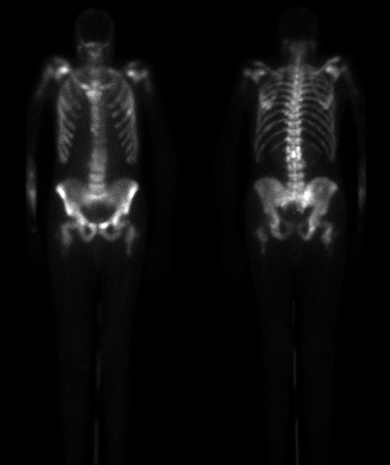
Anterior and posterior images.
View main image(bs) in a separate image viewer
View second image(xr).
Plain film L-spine and pelvis
Full history/Diagnosis is available below
Diagnosis: Metastatic prostate carcinoma
Full history:
92-year old man with known
metastatic prostate carcinoma. The patient had
complaints of diffuse bony pain, most severe in the
back. This examination was obtained to evaluate the
extent of metastases prior to strontium therapy.
Radiopharmaceutical:
Tc-99m MDP
Findings:
Diffuse areas of markedly increased
uptake of the radiopharmaceutical are noted
throughout the axial skeleton. The appendicular
skeleton is relatively spared with the exception of the
proximal aspect of the right forearm. These findings
are most consistent with widespread metastatic
disease. The plain films confirm the presence of
multiple blastic lesions throughout the spine and
pelvis.
Discussion:
Widespread osseous metastatic
disease may sometimes have a superscan appearance.
Key differentiating features from metabolic causes of
superscan include more patchy, heterogeneous bony
uptake as well as the central axial distribution with
relative sparing of the appendicular skeleton.
ACR Codes and Keywords:
References and General Discussion of Bone Scintigraphy (Anatomic field:Skeletal System, Category:Neoplasm, Neoplastic-like condition)
Search for similar cases.
Edit this case
Add comments about this case
Read comments about this case
Return to the Teaching File home page.
Case number: bs054
Copyright by Wash U MO