Case Author(s): Samuel Wang, M.D. and Barry A. Siegel, M.D., 1/12/96 . Rating: #D3, #Q3
Diagnosis: Radiation nephritis
Brief history:
33-year-old woman with history
of breast carcinoma and known osseous metastatic
disease.
Images:

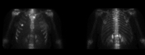
Anterior and posterior images from current study.
View main image(bs) in a separate image viewer
View second image(bs).
Anterior and posterior images performed 10 months previously.
Full history/Diagnosis is available below
Diagnosis: Radiation nephritis
Full history:
33-year-old woman with a history
of breast carcinoma diagnosed 14 months previously
who had undergone a left breast excisional biopsy, 4
courses of chemotherapy, and 1 course of radiation
therapy to the thoracolumbar spine (from the level of
the 8th thoracic vertebra down to and including the
lumbar spine) for osseous metastases. The total radiation
dose to the spine was 3500 cGy, administered over 20 days.
Radiopharmaceutical:
Tc-99m MDP
Findings:
Bone scintigraphy performed 4
months prior to the radiation therapy to the
thoracolumbar spine demonstrated focal areas of
increased activity most consistent with metastases in
the 10th, 11th, and 12th thoracic vertebrae, the 2nd,
3rd, and 4th lumbar vertebrae, and the right 5th and
8th ribs. Repeat bone scintigraphy was performed 6
months after the radiation treatment. There was a
decrease in the intensity of the activity in the known
thoracic and lumbar vertebral metastases. In
addition, the study revealed 2 new focal areas of
increased activity in a linear distribution parallel to
the 12th thoracic vertebra. This new abnormality
appeared separate from the vertebra. Radiographs
demonstrated compression and sclerosis of the
vertebral body without extension into the soft tissues
or soft tissue calcifications. The scintigraphic findings
were felt to represent acute segmental radiation
nephritis involving the superomedial aspects of the
kidneys.
Discussion:
The kidneys are highly
radiosensitive organs. Radiation-induced injury
occurs in proportion to the radiation dose and the
volume of renal tissue exposed. In general, radiation
damage to the kidneys occurs with dosages greater
than 2300 cGy over approximately a five-week period.
Acute radiation nephritis occurs at 6-13 months and
chronic radiation nephritis occurs at 18 months to
several years after treatment. Patients on
chemotherapy have a greater propensity for injury.
This case illustrates the incidental scintigraphic
features of acute segmental radiation nephritis in a
patient who received radiation therapy to the
thoracolumbar spine. The geometric borders of the
area of increased activity correspond to the lateral
limits of the radiation port, which includes the
superomedial aspects of the kidneys. This intensity of
activity on 2-hour delayed images reflects the
abnormal retention of radiotracer within the renal
parenchymal, which has undergone acute radiation
damage.
references:
1) Dewitt, et al.
International J Radiation Oncol and Biology Physics
1990;19:977-983
2) Palestro, et al. Clin Nucl Med
1988;13:789-791
ACR Codes and Keywords:
References and General Discussion of Bone Scintigraphy (Anatomic field:Genitourinary System, Category:Effect of Trauma)
Search for similar cases.
Edit this case
Add comments about this case
Return to the Teaching File home page.
Case number: bs053
Copyright by Wash U MO

