Case Author(s): Charles Pringle, M.D.,Farrokh Dehdashti, M.D. , 02/13/96 . Rating: #D4, #Q4
Diagnosis: Neuroblastoma
Brief history:
Intermittent back pain for three
months.
Images:
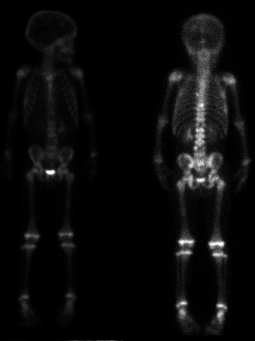
Whole body delay images
View main image(bs) in a separate image viewer
View second image(ot).
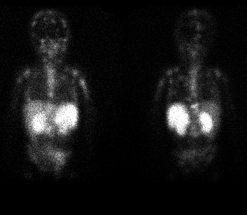
24-hour delay images
View third image(ct).
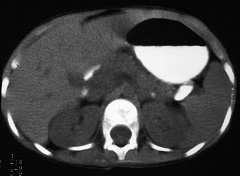
transverse image through upper abdomen
Full history/Diagnosis is available below
Diagnosis: Neuroblastoma
Full history:
5-1/2 year old boy complaining of
worsening, intermittent back pain over the last three
months.
Radiopharmaceutical:
Bone scintigraphy - 4.6
mCi Tc-99m MDP i.v. and Octreotide scintigraphy -
1.4 mCi In-111 octreoscan i.v.
Findings:
The bone scintigraphy demonstrates
multiple focal areas of increased radiopharmaceutical
activity involving the thoracolumbar spine, the skull,
and the left pelvis. Additionally, there are two
photopenic areas present within the T12 vertebra and
within the L4 vertebra. These findings are most
suggestive of malignancy. The differential would
include metastatic disease, lymphoma, or leukemia.
The octreotide scintigraphy demonstrates multiple
focal areas of increased activity throughout the axial
and appendicular skeleton including the skull, the
face, the clivus, the sternum, bilateral proximal
humeri, bilateral ribs, multiple areas within the
entire spine, the pelvis, bilateral femora, and bilateral
proximal tibiae. This appearance is consistent with
somatostatin receptor-positive metastatic disease.
Discussion:
After the initial bone scintigraphy,
this patient underwent a needle biopsy of a lumbar
vertebra. This revealed a small round blue cell tumor.
The most likely diagnosis was metastatic
neuroblastoma. However, initial interpretation of
magnetic resonance imaging and computed
tomography of the chest, abdomen, and pelvis did not
reveal a primary tumor. Therefore, this patient was
scheduled for octreotide scintigraphy to evaluate for a
primary neuroblastoma. As stated above, the
octreotide scintigraphy did confirm the multiple areas
of bony metastatic disease. However, no definite
primary tumor was identified.
References:
Krenning EP et al.
Somatostatin scintigraphy with
Followup:
This patient received a single dose
of cisplatin chemotherapy. Two hours later, the
patient began having opsoclonus and myoclonus. The
patient went into full-blown status epilepticus and
subsequently expired. The exact cause of death was
not determined. However, at autopsy, a 1.5 - 2 cm left
adrenal neuroblastoma was identified.
Major teaching point(s):
Neuroblastoma is the most
common solid abdominal mass of infancy and the
second most common tumor in childhood. These
patients may present with pain and fever, a palpable
abdominal mass, bone pain, or inability to walk.
There is increased catecholamine production in 75-
90%. This is identified often as increased VMA and
HVA in the urine. Octreotide scintigraphy visualized
tumor deposits in about 90% of patients with
neuroblastoma. Patients with somatostatin-receptor-
positive tumor scans have a longer survival rate
compared with receptor-negative patients. These
tumors may also be identified using MIBG. Tumor
visualization is very similar with octreotide and MIBG
scintigraphy.
ACR Codes and Keywords:
References and General Discussion of Bone Scintigraphy (Anatomic field:Genitourinary System, Category:Neoplasm, Neoplastic-like condition)
Search for similar cases.
Edit this case
Add comments about this case
Read comments about this case
Return to the Teaching File home page.
Case number: bs052
Copyright by Wash U MO