Case Author(s): Gregg D. Schubach, M.D. and Jerold W. Wallis, M.D. , . Rating: #D2, #Q5
Diagnosis: Giant Cell Tumor
Brief history:
29-year old man with left knee
pain for several weeks.
Images:
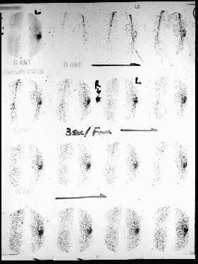
Anterior Flow Images... (upper left image is immediate static/blood pool image)
View main image(bs) in a separate image viewer
View second image(bs).
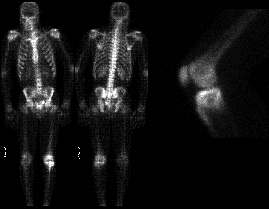
Anterior and Posterior whole body delayed images and spot view left knee
View third image(xr).
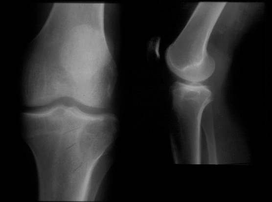
AP and lateral of left knee
View fourth image(mr).
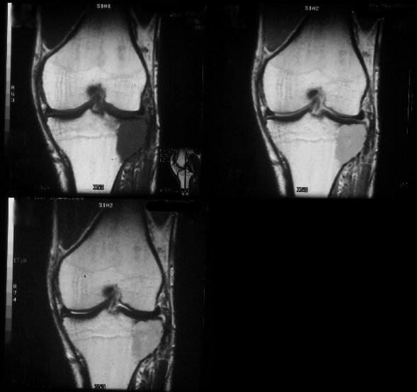
T1, Proton density, and T2 coronal images
Full history/Diagnosis is available below
Diagnosis: Giant Cell Tumor
Full history:
29-year old man with left knee
pain. There is no history of trauma, primary
malignancy, musculoskeletal disorders, or other
pertinent past medical problems. The patient
presents to Nuclear Medicine following plain
radiographs and an MRI examination of the left knee.
Radiopharmaceutical:
Tc-99m MDP i.v.
Findings:
Three-phase bone scintigraphy
demonstrates increased flow and delayed radiopharmaceutical
uptake to the lateral aspect of the left tibial plateau.
The lateral view confirms that the major finding is in the
tibial plateau rather than the patella.
The plain radiograph dated 10-4-95 reveals a lytic
³end-of-bone² lesion along the lateral aspect of the left
tibial plateau. The lesion is characterized by a non-
mineralized matrix, narrow zone of transition, and no
sclerotic rim. The MRI study demonstrates a 2 x 3 x 3
cm mass occupying the lateral aspect of the left tibial
plateau. The lesion is sharply demarcated from the
marrow in its medial aspect, but abuts the cortex
laterally and posteriorly (only coronal images shown).
The lesion is low signal intensity on T1 images and
high signal intensity on T2 weighted images. The
cortex is disrupted through the superior aspect of the
lateral margin of the tibial plateau, posterior margin,
as well as the articular surface. The mass extends
outside of the bone and beneath the posterior horn of
the lateral meniscus. The mass bows the collateral
ligament slightly laterally. The medial collateral,
cruciate, and the patellar ligaments appear intact.
Discussion:
Giant cell tumors, which are also
called osteoclastomas, account for approximately 4%
of all primary bone tumors and approximately 21% of
all benign skeletal tumors. Giant cell tumors affect
men and women with equal frequency and typically
occur between ages 20 and 40 years. This lesion is
rare in children. Giant cell tumors are typically
eccentric ³end-of-bone² lesions seen in long bones with
50-60% about the knee. The lesion is characterized by
a non-mineralized matrix, narrow zone of transition,
and no sclerotic rim. 25% demonstrate soft tissue
invasion and 5% are complicated by a pathologic
fracture. Uptake on bone scintigraphy is typically intense,
and may extend slighlty beyond the limits of the tumor.
85% of the giant cell tumors are benign.
However, the giant cell tumor is a
neoplasm which, although appearing histologically benign, may
metastasize. Case reports in the recent literature
describe metastatic lung nodules which, upon surgical
resection, demonstrate giant cell lesions with clearly
benign histopathology. Thus, the surgical resection of
the ³mets², along with treatment of the primary lesion
(usually by curettage and grafting) is considered
curative. The rate of recurrence of a giant cell tumor
is about 40-60% with most recurrences occurring
within two years after treatment and usually at the
site of the primary tumor.
Bone scintigraphy was performed primarily to look for additional
bone lesions. Three-phase
(rather than single-phase) scintigraphy was performed for
academic interest at no
additional cost to the patient.
References: Resnik. Bone and Joint
Imaging. 1989
Followup:
The patient underwent curettage of
the lesion with bone grafting and/or packing with
bone chips.
Major teaching point(s):
1) Many bone abnormalities,
including neoplasms, are ³hot² on three-phase bone
scintigraphy. Most neoplasms demonstrate increased
vascularity; hence, there is increased flow on the
scintigraphic angiogram. Demonstrating this is of no
additional diagnostic value; however, it is useful to
note that tumor remains in the differential when
three-phase
scintigraphy is used for other reasons (e.g. to assess
for osteomyelitis).
2) Giant cell tumors have a classic radiographic
appearance including; a) an ³end-of-bone² lesion, b) no
sclerotic rim, and c) no mineralized matrix.
3) Specific age range at presentation (20-40 yrs) is
helpful in distinguishing similarly appearing lesions.
Differential Diagnosis List
The classic
³end-of-bone² lesions include: 1) Giant cell tumor; 2)
Chondroblastoma; 3) Intraosseous ganglion; 4)
Chondrosarcoma (clear cell); and 5) Paget¹s disease.
Chondroblastoma typically occurs in a child or
adolescent and may contain chondroid calcifications.
The intraosseous ganglion is observed most frequently
in the medial malleolus of the tibia, in the carpal
bones, or the periarticular regions such as the hip.
The radiographic appearance and age at presentation
are characteristic of giant cell tumor. If multiple
giant cell tumors are being considered, brown tumors
(seen in hyperparathyroidism) should be considered.
Based on the scintigraphic appearance alone, the differential
would include tumor, fracture, healing avascular necrosis, and
osteomyelitis.
ACR Codes and Keywords:
References and General Discussion of Bone Scintigraphy (Anatomic field:Skeletal System, Category:Neoplasm, Neoplastic-like condition)
Search for similar cases.
Edit this case
Add comments about this case
Read comments about this case
Return to the Teaching File home page.
Case number: bs047
Copyright by Wash U MO