Case Author(s): Gregg D.Schubach, M.D. and Keith C. Fischer, M.D. , . Rating: #D3, #Q5
Diagnosis: Pachydermoperiostosis
Brief history:
45-year old man with diffuse
aches, pains, and arthralgias.
Images:

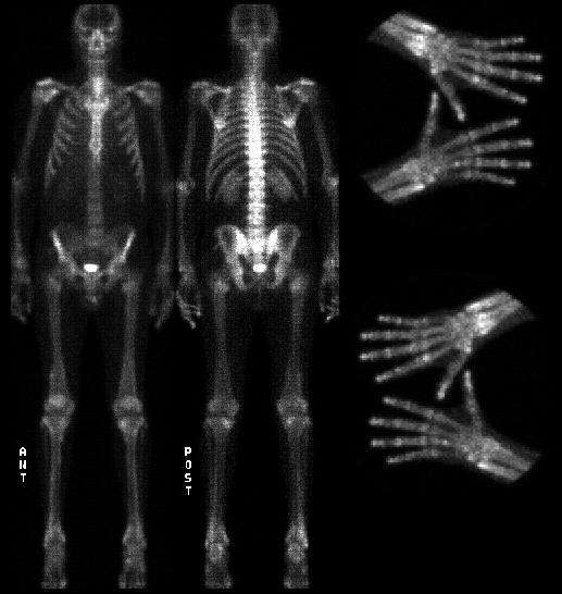
Anterior and posterior delayed whole body images and spot views of the hands.
View main image(bs) in a separate image viewer
View second image(xr).
Anterior view of femur
View third image(xr).
AP view of hand
View fourth image(xr).
Anterior view of tibia
Full history/Diagnosis is available below
Diagnosis: Pachydermoperiostosis
Full history:
45-year old man with a diagnosis
of pachydermoperiostosis diagnosed at age 24. The
patient currently presents with diffuse achiness and
arthralgias. On physical examination, the patient has
marked clubbing of the fingers, skin thickening
(hyperkeratosis) of the palms of the hands and soles of
the feet, as well as grossly enlarged joints at the
wrists, knees, and ankles. Laboratory data is normal
except for an elevated erythrocyte sedimentation rate.
The patient has had multiple tendon release
operations as treatment for joint pain. The patient is
currently being treated with analgesics as needed.
Radiopharmaceutical:
Tc-99m MDP
Findings:
The delayed whole body bone
scan demonstrates enlargement of the distal
diaphyses and metaphyses of the femora. There is
mildly increased radiopharmaceutical uptake along
the periosteum of the distal femora, tibiae, and
forearms bilaterally. There is also increased activity
in the hands and feet as well as several other foci.
These scintigraphic abnormalities correspond to the
periosteal reaction noted on the plain radiographs.
The plain radiographs demonstrate diffuse periosteal
reaction about the distal femora, tibiae, radii, and
spade-like tufts of the fingers.
Discussion:
Hypertrophic osteoarthropathy
may be divided into primary (also called hereditary or
idiopathic) and secondary. The primary form, which
accounts for approximately 5% of all cases of
hypertrophic osteoarthropathy, is also called
pachydermoperiostosis. Secondary hypertrophic
osteoarthropathy, also called pulmonary hypertrophic
osteoarthropathy, is secondary to a variety of
underlying cardiopulmonary diseases, such as
bronchogenic carcinoma, mesothelioma, pulmonary
abscess, emphysema, bronchiectasis, Hodgkin¹s
disease, cyanotic congenital heart disease, cystic
fibrosis, etc. Pachydermoperiostosis, which is
inherited as an autosomal dominant, affects
men much more frequently than women. This
disease, which has a predilection for blacks, is
characterized clinically by an insidious onset during
adolescence of enlargement of the hands and feet
and clubbing of the fingers and toes. Patients also
suffer large skin folds on the face and scalp, excessive
perspiration, enlargement of the extremities, and
nonspecific pain in the bones and joints.
Pachydermoperiostosis usually ceases after ten years
of progression. Prior to the spontaneous arrest of
progression, patients may suffer chronic disabling
complications including restricted motion, severe
kyphosis, and neurologic manifestations. Life
expectancy, however, is normal. Radiographic
abnormalities include enlargement of the paranasal
sinuses, irregular periosteal proliferation of the
phalanges and distal long bones beginning at the
epiphyseal regions, cortical thickening without
narrowing of the medullary cavity, clubbing, and
occasional acro-osteolysis. Hypertrophic
osteoarthropathy has a distinctive scintigraphic
appearance. There is increased radiopharmaceutical
uptake along the cortical margins of the diaphyses of
long bones. The juxta-articular regions of the long
bones may be involved, especially in the phalanges.
The bone scan, which is asymmetric or
irregular in about 15% of cases, shows frequently involvement
of the scapulae, mandible, maxilla, clavicles, and patellae.
The scintigraphic findings often precede the
radiographic abnormalities. Specifically,
radiopharmaceutical uptake may be increased
secondary to hyperemia prior to periosteal new bone
formation. Also, nuclear imaging of the hands and
feet may show increased activity in the terminal
phalanges prior to the development of clinically
evident clubbing.
References:
1. Resnik. Bone and Joint Imaging, 1989
2. Wagner, et al. Principles of Nuclear Medicine, 2nd
edition, 1995
Followup:
The family history reveals the
patient¹s father, brother, and sister suffer from this
condition. The patient has two brothers who are not
affected. The patient has four young children without
clinical evidence of pachydermoperiostosis at this
time.
Major teaching point(s):
Hypertrophic
osteoarthropathy should not be, and rarely is,
confused with metastatic disease.
Differential Diagnosis List
The differential
diagnosis for periosteal reaction includes trauma, infection
(osteomyelitis), inflammatory disease (e.g., arthritis),
neoplasm, congenital disorders (physiologic in the newborn),
metabolic
ACR Codes and Keywords:
References and General Discussion of Bone Scintigraphy (Anatomic field:Skeletal System, Category:Organ specific)
Search for similar cases.
Edit this case
Add comments about this case
Read comments about this case
Return to the Teaching File home page.
Case number: bs046
Copyright by Wash U MO

