Case Author(s): Michael Roarke, MD and Jerold Wallis, MD , 11/2/95 . Rating: #D2, #Q4
Diagnosis: Radiation induced osteosarcoma of lumbar spine.
Brief history:
71-year old gentleman with
back pain.
Images:

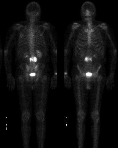
Views from delayed whole body bone scintigraphy.
View main image(bs) in a separate image viewer
View second image(xr).
Anteroposterior view of the lumbar spine.
View third image(ct).
Axial CT image lumbar spine.
Full history/Diagnosis is available below
Diagnosis: Radiation induced osteosarcoma of lumbar spine.
Full history:
71-year old gentleman with a
history of radiation treatment to the abdomen 11
years ago for a lymphoma. The patient recently
developed back pain and radiographs demonstrated a
large osteoblastic lesion of L3.
Radiopharmaceutical:
21.0 mCi Tc-99m MDP
i.v.
Findings:
The bone scintigraphy reveals a
large focus of increased activity corresponding to the
L3 vertebra and extending laterally into the
paravertebral space on each side of the vertebra. No
other suspicious lesions are identified in the skeleton.
Computed tomography examination of the
thoracolumbar spine dated 10-31-95 reveals an
aggressive blastic expansile lesion of the L3 vertebra,
consistent with osteosarcoma.
Discussion:
Approximately 2% of all
osteosarcomas occur in the spine and approximately
4% of all osteosarcomas are radiation induced. Most
osteosarcomas (80%) occur in the appendicular skeleton. This
malignancy is associated with radionuclide treatment
(e.g., following P-32 therapy) as well as external beam
radiation (usual dose greater than 3,000 cGy in three
weeks with a threshold of about 1,000 cGy). The
latency is 4-42 years with an average of 11 years.
Osteosarcomas and malignant fibrous histiocytomas
are the two most common radiation-induced sarcomas.
In a patient with treated Hodgkin¹s disease, the risk
in five-year survivors for radiation-induced
malignancy is approximately 0.9%. There is a
bimodal age distribution for osteosarcoma with the
first peak occurring at 10-25 years old and the second
peak occurring greater than 60 years of age. The
latter is usually associated with pre-existing
conditions such as Paget¹s disease, radiation therapy,
multiple hereditary exostoses, and fibrous dysplasia.
References: Resnick and Niwayama.
Diagnosis of Bone and Joint Disorders, 3rd edition.
ACR Codes and Keywords:
References and General Discussion of Bone Scintigraphy (Anatomic field:Spine and Contents, Category:Neoplasm, Neoplastic-like condition)
Search for similar cases.
Edit this case
Add comments about this case
Read comments about this case
Return to the Teaching File home page.
Case number: bs043
Copyright by Wash U MO

