Case Author(s): Charles Pringle, M.D., Mike Roarke, M.D., and Keith Fischer, M.D. , 11/16/95 . Rating: #D3, #Q4
Diagnosis: Maffucci's syndrome
Brief history:
History witheld.
Images:
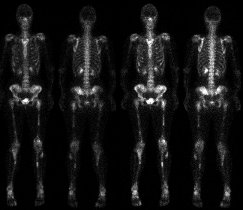
Anterior and posterior delayed images in
two intensity settings.
View main image(bs) in a separate image viewer
View second image(bs).
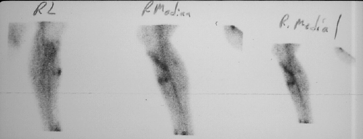
Delayed images of the right lower extremity
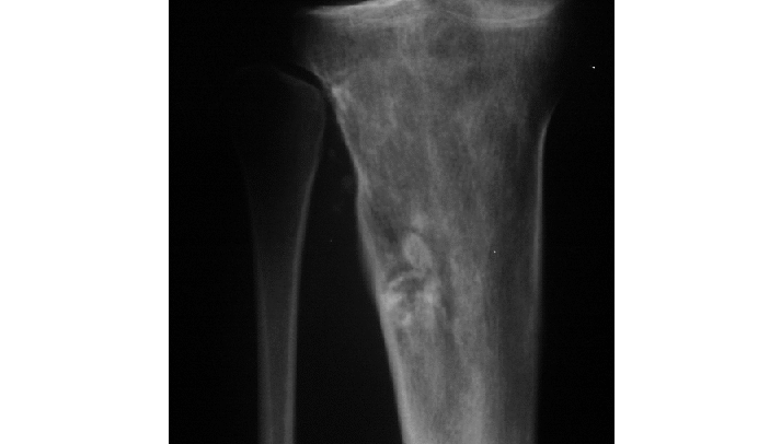
View third image(xr).
Anterior view right tibia and fibula
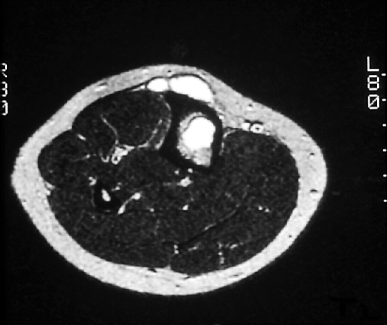
View fourth image(mr).
single axial T2 weighted image through the
proximal tibia and fibula
Full history/Diagnosis is available below
Diagnosis: Maffucci's syndrome
Full history:
26-year old woman with history
of Maffucciąs syndrome (nonhereditary
enchondromatosis and multiple soft tissue cavernous
hemangiomas), for routine follow-up examination.
Radiopharmaceutical:
20.7 mCi Tc-99m MDP
Findings:
There is increased activity in the
soft tissues of the lower extremities on the immediate
images consistent with hemangiomas in these areas.
See plain films. On delayed images, multiple focal
areas of abnormal tracer accumulation throughout the
skeleton, not significantly changed since the previous
study of 4-19-94 (not shown). These lesions
correspond to multiple enchondromata. Also, bowing
of bilateral forearms and lower legs, without change.
Discussion:
This patient undergoes yearly
follow-up examinations to evaluate for malignant
transformation of the enchondromata.
References: Donald Resnick, M.D.
Bone and Joint Imaging. 1989
Followup:
None
Major teaching point(s):
Maffucciąs syndrome is a
nonhereditary, rare cartilaginous dysplasia resulting
in multiple enchondromas and soft tissue
hemangiomas. A unilateral skeletal distribution is
present in approximately 50% of cases. The
metacarpals and phalanges of the hands are most
frequently involved. When the extremities are
involved, there may be resultant limb length
discrepancies. Malignant transformation is the most
severe complication and generally occurs in adults
after the age of 40 years. The approximate frequency
is 20%. There is potential for sarcomatous
degeneration of both bone and soft tissue lesions.
Chondrosarcoma is the most common, but other
reported neoplasms include hemangiosarcoma,
lymphangiosarcoma, and fibrosarcoma.
Differential Diagnosis List
Based on the
bone scintigraphy alone, without any history, a
differential diagnosis would include metastases or a
metabolic bone disease.
ACR Codes and Keywords:
References and General Discussion of Bone Scintigraphy (Anatomic field:Skeletal System, Category:Normal, Technique, Congenital Anomaly)
Search for similar cases.
Edit this case
Add comments about this case
Read comments about this case
Return to the Teaching File home page.
Case number: bs042
Copyright by Wash U MO