Case Author(s): Gregg D. Schubach, MD and Tom R. Miller, MD, PhD , 10/13/95 . Rating: #D3, #Q4
Diagnosis: Hypophosphatemia/X-linked Vitamin D-resistant Rickets (osteomalacia)
Brief history:
25-year old woman with
complaints of low back and left knee pain for one
year.
Images:

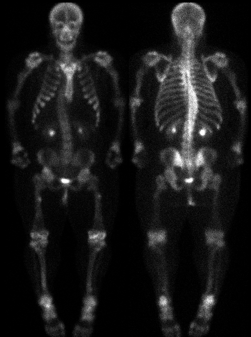
Anterior and Posterior Whole Body Images
View main image(bs) in a separate image viewer
View second image(bs).
Lateral images of the left and right knee, respectively.
View third image(xr).
Radiographs of the left knee
View fourth image(xr).
Spot image of the proximal left femur
Full history/Diagnosis is available below
Diagnosis: Hypophosphatemia/X-linked Vitamin D-resistant Rickets (osteomalacia)
Full history:
25-year old woman with a
history of vitamin D-resistant rickets. The patient has undergone spinal fusion and multiple
osteotomies to the lower extremities bilaterally.
The most recent surgery was approximately nine
years ago. The patient now complains of low back
and left knee pain for one year.
Radiopharmaceutical:
Tc-99m MDP
Findings:
Bone scintigraphy demonstrates
focal regions of increased radiopharmaceutical
uptake in the proximal right femoral diaphysis
medially and the left proximal femoral diaphysis
laterally. This corresponds to the Looser's
zones (pseudofractures) on the plain radiographs of
the femora. There is scintigraphic evidence of
Harrington rods in the thoracolumbar region in this
patient with a lower thoracic dextroscoliosis and
short stature. Note is also made of the suboptimally
positioned gonadal shield on the plain radiographs.
There is shortening and bowing deformities of
multiple long bones, including the humeri and
femora, which is corroborated on the plain
radiographs of the knees. No scintigraphic
abnormality is identified in the distal left femur,
proximal left tibia, or proximal left fibula to
correspond with the foci of periosteal reaction on
the radiographs. The radiographic abnormalities,
some of which have scintigraphic correlates, likely
represent Looser1s zones in various phases of
healing. The short stature, bone deformities, and
Looser1s zones are characteristic of osteomalacia.
Discussion:
Several rachitic and
osteomalacic syndromes, which share one or several
renal tubular abnormalities, are collectively known
as Fanconi syndromes. X-linked hypophosphatemia
(also known as familial vitamin D-resistant rickets)
is the most common form of renal tubular rickets
and osteomalacia. This syndrome, which is
characterized by life-long hypophosphatemia
secondary to renal tubular phosphate loss, decreased
intestinal absorption of calcium, and normal serum
levels of calcium, usually appears between 12 and
18 months of age. Afflicted individuals are short,
bowlegged, stocky, and usually men (X-linked
dominant trait). The development and severity of
rachitic changes differ from patient to patient and
systemic symptoms, such as muscle weakness and
hypotonia, are absent. Although remission typically
follows growth plate closure, recurrence of
symptoms is common in adulthood. Radiographic
features include bowing of long bones (especially
lower extremities), rachitic changes at the growth
plates, and minimal osteopenia. Coarsening of the
trabecular pattern along with Looser1s zones are
identified with aging. The latter may be
complicated by complete fractures. The skeleton
shows multiple sites of new bone formation at
various tendinous and ligamentous attachments.
These enthesopathic calcifications/ossifications may
be identified in the annulus fibrosis, paravertebral
ligaments, apophyseal joints, acetabulum,
iliolumbar ligaments, and sacroiliac ligaments.
Also, periarticular ossicles may develop as small
separate entities, especially about the carpus.
Treatment of hypophosphatemia includes phosphate
infusion and large doses of vitamin D.
References: Resnik D. Bone and
Joint Imaging.
Major teaching point(s):
The short stature, bone deformities, and
Looser's zones are characteristic of osteomalacia.
ACR Codes and Keywords:
References and General Discussion of Bone Scintigraphy (Anatomic field:Skeletal System, Category:Metabolic, endocrine, toxic)
Search for similar cases.
Edit this case
Add comments about this case
Read comments about this case
Return to the Teaching File home page.
Case number: bs041
Copyright by Wash U MO

