Case Author(s): M.Roarke, M.D., F. Vanwagenen, M.D., J. Wallis, M.D. , 10/6/95 . Rating: #D3, #Q5
Diagnosis: Diffuse Paget's Disease
Brief history:
Painful palpable mass in the right anterior iliac
region.
Images:
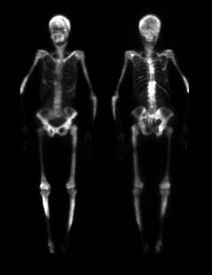
Anterior and posterior images from whole body delayed bone scintigraphy.
View main image(bs) in a separate image viewer
View second image(xr).
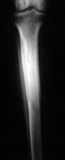
AP radiograph of the right tibia.
View third image(xr).
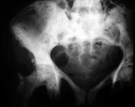
AP pelvis radiograph.
View fourth image(mr).
Pelvis MRI.
Full history/Diagnosis is available below
Diagnosis: Diffuse Paget's Disease
Full history:
45-year old black woman with
known neurofibromatosis 1 and history of Paget¹s
disease diagnosed three years ago. She now presents
with a painful palpable mass in the right anterior iliac
region. The alkaline-phosphatase measured 400.
This examination was requested to evaluate for
evidence of scintigraphic abnormality associated with
the palpable mass as well as to confirm the diagnosis
of Paget¹s disease. There is no history of metabolic
bone disease, metastatic disease, primary malignancy
or fibrous dysplasia.
Radiopharmaceutical:
21.8 mCi Tc-99m MDP
i.v.
Findings:
There are multiple areas of
markedly increased tracer uptake throughout the
skeleton; findings at several sites, such as the tibiae and
right hemipelvis (most evident on the posterior image
in the peri-acetabular region), suggest Paget¹s disease. However,
many of the sites would be difficult to distinguish
from diffuse metastatic disease. Plain radiographs of
the areas of abnormality on scintigraphy reveal the
characteristic findings in Paget¹s disease. In the spine
and pelvis, sclerosis, coarsening of trabeculae, and
slight enlargement of bone are all findings consistent
with the sclerotic phase of Paget¹s disease. In the
tibiae, an advancing front of lytic change is present
radiographically, giving the so-called ³blade of grass²
appearance of the lytic phase of Paget¹s disease. The
pelvic MRI demonstrated multiple masses which were
hyperintense on T-2 weighted sequences and which
enhanced with i.v. gadolinium administration.
Discussion:
Paget¹s disease is common,
occurring in 3.5% of the population greater than 40
years old and in 10% of the population greater than 80
years old. The onset of the disease in the population
less than 40 years old is uncommon. The etiologic
agent is likely a paramyxovirus. The phases of
Paget¹s disease include osteoclastic,
osteoblastic/clastic, and osteosclerotic or quiescent
phases. Malignant transformation represents a
fourth phase. 85% of cases are polyostotic. Clinical
features include pain, fractures, nerve compression,
skeletal deformities, and osteoarthritis. Laboratory
analysis reveals elevated alkaline-phosphatase and
urinary hydroxyproline. Paget¹s disease is associated
with sarcomatous transformation, giant cell tumors,
plasma cell myeloma, and metastases. In this case,
the right iliac soft tissue mass demonstrated on
magnetic resonance imaging examination was
biopsied and proven to represent a giant cell tumor of
the tendon sheath rather than a malignancy such as a
sarcoma.
References: Resnick, Niyawma.
Diagnosis of Bone and Joint Disorders, 3rd edition.
Major teaching point(s):
Comparison with plain
radiographs of scintigraphically abnormal regions is
frequently necessary for diagnosis.
Differential Diagnosis List
The
differential diagnosis includes metastatic disease, Paget's
disease, and
polyostotic fibrous dysplasia.
ACR Codes and Keywords:
References and General Discussion of Bone Scintigraphy (Anatomic field:Skeletal System, Category:Neoplasm, Neoplastic-like condition)
Search for similar cases.
Edit this case
Add comments about this case
Read comments about this case
Return to the Teaching File home page.
Case number: bs040
Copyright by Wash U MO