Case Author(s): J. Philip Moyers, MD and Jerold Wallis, M.D. , 10/3/95 . Rating: #D2, #Q3
Diagnosis: Prostate Carcinoma, metastatic to liver
Brief history:
Prostate carcinoma
Images:
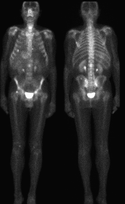
Anterior and posterior whole body delayed images
View main image(bs) in a separate image viewer
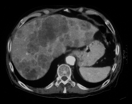
View second image(ct).
Single axial image from a contrast enhanced CT of the abdomen
Full history/Diagnosis is available below
Diagnosis: Prostate Carcinoma, metastatic to liver
Full history:
69-year old man with known
prostatic carcinoma diagnosed in March 1992.
Radical prostatectomy was performed in June 1992.
The patient is currently under treatment with
leuprolide (Lupron®), and has otherwise not had
chemotherapy. The patient has known hepatic and
osseous metastatic disease.
Radiopharmaceutical:
22.0 mCi Tc-99m MDP i.v.
Findings:
Multiple areas of increased uptake
consistent with osseous metastases are demonstrated,
including the right femoral neck, L2, the left scapula,
and several sites in the pelvis. Also noted is an
abnormal focus of increased activity in the right upper
quadrant of the abdomen. Correlation with the CT
scan dated 8-17-95 demonstrates diffuse hepatic
metastases. A single very large hepatic metastasis is
demonstrated in the medial segment of the left
hepatic lobe, which likely corresponds to the abnormal
focus of increased soft tissue tracer uptake.
Discussion:
There are many causes of
abnormal soft tissue uptake on bone scintigraphy.
Bone scintigraphy agents have historically included a variety of
Tc-99m labeled
phosphate and diphosphonate compounds, with current use of
methylene diphosphonate (MDP) and
hydroxymethylene diphosphonate (HMDP). Like the early agent
pyrophosphate (still used for myocardial infarct imaging), these
radiopharmaceuticals tend to localize in areas of
dystrophic calcification or necrotic tissues. Metastatic
adenocarcinomas from ovarian, breast and GI
malignancies commonly undergo necrosis and develop
dystrophic calcification.
References:
1) Mettler FA.
Essentials of Nuclear Medicine Imaging. 1991, 3rd
edition.
2) Datz FL. Handbook of Nuclear Medicine, Mosby
Yearbook Publishers, 1993, 2nd edition.
Followup:
CT scan dated 8-17-95
demonstrates diffuse hepatic metastatic disease.
Major teaching point(s):
Evaluation of soft tissue
uptake on bone scintigraphy can sometimes be a clue
in evaluation of disease processes. In this case,
abnormal uptake is demonstrated within the axial
skeleton as well as the right upper quadrant. If one
attributes both findings to a single pathologic process,
the diagnosis of metastatic disease involving the liver
and bone becomes highly likely.
ACR Codes and Keywords:
References and General Discussion of Bone Scintigraphy (Anatomic field:Skeletal System, Category:Neoplasm, Neoplastic-like condition)
Search for similar cases.
Edit this case
Add comments about this case
Read comments about this case
Return to the Teaching File home page.
Case number: bs039
Copyright by Wash U MO