Case Author(s): J. Philip Moyers, MD , 9/30/95 . Rating: #D2, #Q4
Diagnosis: Osteonecrosis, right lateral femoral condyle
Brief history:
Worsening right knee pain.
Images:
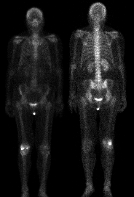
Anterior and posterior whole body bone images
View main image(bs) in a separate image viewer
View second image(bs).
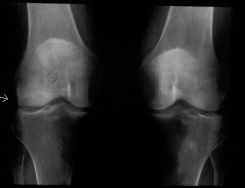
Lateral and medial views of the right knee
View third image(xr).
Current plain films of the knees, AP view
View fourth image(xr).
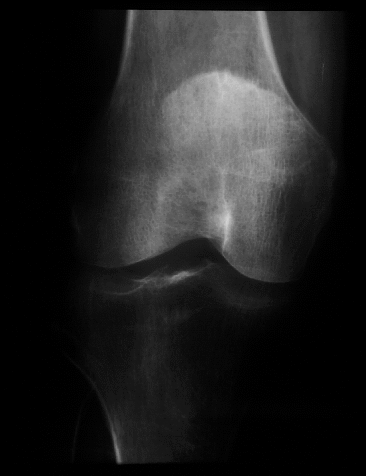
Anterior view of the right knee, approximately 10 months previous
Full history/Diagnosis is available below
Diagnosis: Osteonecrosis, right lateral femoral condyle
Full history:
72-year old woman who had
medial and lateral meniscectomy of the right knee by
arthroscopic surgery. The patient developed acute onset of
pain six months later with increasing difficulty
walking since that time. Plain films were obtained for
evaluation followed by bone scintigraphy.
Radiopharmaceutical:
21.6 mCi Tc-99m MDP
i.v.
Findings:
Bone scintigraphy demonstrates
increased activity in both the medial and lateral
condyles of the right femur. The activity in the lateral
femoral condyle is greater than that in the medial
condyle. Otherwise, no abnormal uptake is
demonstrated throughout the remainder of the
skeleton. Comparison with the bone radiographs
obtained the same day, as well as comparison with a
prior pre-operative radiograph demonstrates an area
of sclerosis and flattening in the lateral femoral
condyle with an osteochondral defect. This is a
dramatic change from the normal pre-operative
radiographs. In this patient population with this
history, findings of osteonecrosis are suggested.
Discussion:
This patient had no pre-disposing
risk factors for avascular necrosis. Osteonecrosis is
usually seen in older patients and is distinct from
osteochondritis dissecans, which occurs in
adolescence. The vast majority of patients
demonstrate changes involving the weight bearing
surfaces of the medial femoral condyle with
involvement of the lateral femoral condyle seen less
frequently. Meniscal tears have been reported in
association with this pathologic entity. This patient
had a meniscectomy six months earlier.
Reference: Resnick D. Bone and
joint imaging. WB Saunders & Co. 1989
Followup:
Plain film correlation of the bone
scintigraphy suggests osteonecrosis involving the
lateral femoral condyle of the right femur.
Major teaching point(s):
Increased activity in the
lateral femoral condyle is demonstrated in the same
region as sclerosis on the plain films. However,
increased activity also is demonstrated in the medial
femoral condyle, which likely represents
subradiographic osteonecrosis.
ACR Codes and Keywords:
References and General Discussion of Bone Scintigraphy (Anatomic field:Skeletal System, Category:Effect of Trauma)
Search for similar cases.
Edit this case
Add comments about this case
Read comments about this case
Return to the Teaching File home page.
Case number: bs038
Copyright by Wash U MO