Case Author(s): J. Philip Moyers, MD , 10/2/95
. Rating: #D2, #Q4
Diagnosis: Soft tissue abscess involving left superior and inferior pubic ramus
Brief history:
Decreased ambulation on the
left lower extremity.
Images:

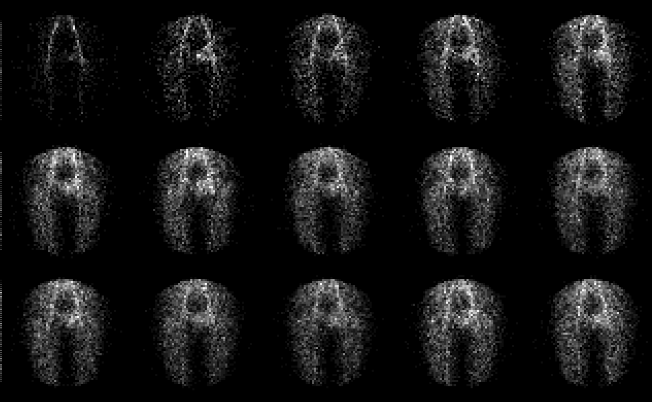
Anterior flow images of the pelvis
View main image(bs) in a separate image viewer
View second image(bs).
Anterior delayed images of the pelvis
View third image(xr).
Anterior plaim film of the pelvis
View fourth image(mr).
Axial MR images and intraoperative ultrasound image of the same area
Full history/Diagnosis is available below
Diagnosis: Soft tissue abscess involving left superior and inferior pubic ramus
Full history:
Patient had prior documented E
coli urinary tract infection four weeks before this
study. The patient currently presents with a 1-3 week
history of decreased ambulation of the left lower
extremity. The patient currently has an increased
white cell count and a moderately increased
sedimentation rate. Plain film of the pelvis suggests
an obstruction in the inferior pubic ramus medial to
the ischial pubis synchondrosis. The patient was
referred for three-phase bone scintigraphy to evaluate
for osteomyelitis.
Radiopharmaceutical:
5.1 mCi Tc-99m MDP
i.v.
Findings:
The anterior radionuclide
angiogram demonstrates increased flow in the region
of the left groin. Immediate static images also
demonstrate increased activity in the left superior and
inferior pubic region. The combination of findings of
increased flow and a destructive lesion in the left
inferior pubic ramus prompted an MRI of the pelvis,
which demonstrates abnormal tissue which enhances
in the left obturator internus. At this time, soft tissue
infection with involvement of bone marrow was
suggested, but differential diagnosis included
rhabdomyosarcoma, Ewingıs tumor, or teratoma. The
patient had an endovaginal ultrasound which
demonstrated a complex hyperechoic mass in this
region.
Discussion:
Three-phase bone scintigraphy
includes radionuclide angiogram over the region of
interest followed by immediate static images and
delayed static images approximately 2-3 hours after
the initial injection of radiopharmaceutical. These
three-phase scintigraphic techniques improve the
specificity of bone scintigraphy. The differential is
between osteomyelitis vs cellulitis. However, if soft
tissue inflammation is a prominent feature, primary
bone involvement may be difficult to distinguish from
increased activity in the bone secondary to hyperemia
from an overlying simple cellulitis. Possible false-
negative studies include an early stage of the disease.
Further evaluation for osteomyelitis can be obtained if
osteomyelitis is strongly clinically suspected with
gallium-67 scintigraphy. If the uptake of gallium in
the region of suspected osteomyelitis is greater than
the uptake of the bone agent, osteomyelitis is
probable. Acute hematogenous osteomyelitis occurs
more commonly in children than in adults. These
cases usually occur in the metaphysis of the long
bones. This is felt to be secondary to the vascular
anatomy in the metaphysis of the long bones.
Children older than 1 year, but prior to skeletal
maturity have no vessels crossing the physeal growth
plates. Vessels approaching the physis from the
nutrient foramen in the diaphysis of the long bones
ramify at the level of the physeal growth plates and
undergo a 180 degree bend back towards the
diaphysis. At this point, there is turbulence of blood
flow, which is postulated to be a cause for the
increased incidence of osteomyelitis in the
metaphysis. In children less than 1 year of age, a
feeding vessel can span the physis and epiphyseal
osteomyelitis occurs with a greater frequency in this
age group. Sometimes, an antecedent history of
trauma can be elicited. The antecedent history of
trauma is postulated to be a predisposing factor for
osteomyelitis by marrow edema in the end of the
bones and an increase in pressure of the marrow
space which results in decrease in blood flow in this
region. The usual findings of osteomyelitis are
increased activity in blood flow, blood pool, and
delayed images. However, cold defects can be
demonstrated due to increased pressure in the
marrow space reducing blood flow, stripping away of
periosteum due to pus, and interruption of blood
supply by sludge and thrombosis of the nutrient
vessels.
References: 1) Mettler FA.
Essentials of Nuclear Medicine Imaging. 1991, 3rd
edition. 2) Datz FL. Handbook of Nuclear
Medicine, Mosby Yearbook Publishers, 1993, 2nd
edition.
Followup:
The patient underwent
intraoperative ultrasound in the operating room
under anesthesia and transperineal biopsy of the
hyperechoic lesion demonstrated by the endovaginal
ultrasound. Several passes were made in this patient
and findings of necrotic abscess were demonstrated.
The patient was placed on antibiotics with prompt
improvement of symptoms.
Major teaching point(s):
The three-phase bone
scintigraphy identified increased activity in the
superior pubic ramus although the inferior pubic
ramus was the area questioned on the plain
radiographs. Abnormal marrow signal is
demonstrated in both the inferior and superior pubic
rami on the MRI study. The patient had a
documented E coli infection approximately four
weeks prior to the bone scintigraphy. Cultures of
the abscess demonstrated Staphylococcus aureus,
which is the most common cause of osteomyelitis.
Differential Diagnosis List
Soft tissue
tumor such as rhabdomyosarcoma or Ewingıs
sarcoma might also show increased activity on all
three phases of three-phase bone scintigraphy.
ACR Codes and Keywords:
References and General Discussion of Bone Scintigraphy (Anatomic field:Skeletal System, Category:Inflammation,Infection)
Search for similar cases.
Edit this case
Add comments about this case
Read comments about this case
Return to the Teaching File home page.
Case number: bs037
Copyright by Wash U MO

