Case Author(s): M. Roarke , 9/1/95 . Rating: #D2, #Q4
Diagnosis: Osteitis Condensans of the Clavicle
Brief history:
33 year-old woman with a three month history of left medial clavicular pain.
Images:

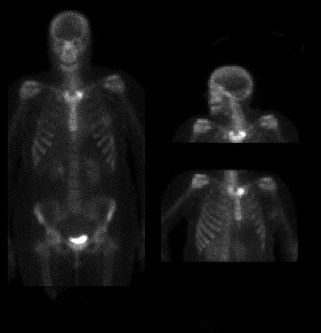
Images from whole body bone scintigraphy.
View main image(bs) in a separate image viewer
View second image(ct).
Transaxial CT image at the claviculomanubrial articulation.
Full history/Diagnosis is available below
Diagnosis: Osteitis Condensans of the Clavicle
Full history:
33 year-old woman with three month history of left medial clavicular pain. There was no history of known trauma, fever, arthritis or intravenous drug abuse.
Radiopharmaceutical:
Tc-99m MDP i.v.
Findings:
Moderately increased focal uptake is seen in the medial ends of both clavicles, left greater than right. The computed tomographic examination confirmed sclerosis in the clavicular heads, left greater than right. These findings, coupled with the clinical presentation, are most consistent with osteitis condensans of the clavicles.
Discussion:
Osteitis condensans of the clavicles tends to occur in women with an average age of 40 years. It is characterized clinically by pain and swelling over the medial end of the clavicle. Bony sclerosis and eburnation may occur, but the sternoclavicular joint remains intact. The etiology of this disorder is unclear, but it may represent a response to mechanical stress. Differential diagnostic possibilities include ischemic ncecrosis of the medial clavicular epiphysis, sternocostoclavicular hyperostosis and septic arthritis of the sternoclavicular joint. Clinical information usually helps to distinguish among these possibilities.
ACR Codes and Keywords:
References and General Discussion of Bone Scintigraphy (Anatomic field:Skeletal System, Category:Effect of Trauma)
Search for similar cases.
Edit this case
Add comments about this case
Read comments about this case
Return to the Teaching File home page.
Case number: bs034
Copyright by Wash U MO

