Case Author(s): Samuel Wang, M.D., Tom R. Miller, M.D., Ph.D. , 9/22/95 . Rating: #D3, #Q3
Diagnosis: Neuroblastoma and spiral fracture of the right femur
Brief history:
8-month old boy who presented
initially with constipation
Images:

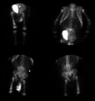
Multiple spot images.
View main image(bm) in a separate image viewer
View second image(gu).
Cystogram.
View third image(ct).
CT of pelvis.
View fourth image(xr).
Radiograph of femur.
Full history/Diagnosis is available below
Diagnosis: Neuroblastoma and spiral fracture of the right femur
Full history:
8-month old boy who presented
initially with constipation and a palpable pelvic and
lower abdominal mass. The patient was diagnosed
with stage IV neuroblastoma with extension into the
spinal canal. The patient had undergone an L3-L5
laminectomy and was receiving chemotherapy at the time of the bone scan.
Bone scintigraphy performed two months prior to the present
study had demonstrated the mass, but no evidence of
osseous metastases.
Radiopharmaceutical:
Tc-99m MDP i.v.
Findings:
Bone scintigraphy demonstrates
increased uptake of the radiopharmaceutical in the
soft tissues of the pelvis. The bladder is displaced to
the right. These findings correspond to the site of the
patient's known neuroblastoma, which contains foci of
calcifications as seen on the CT study and on
cystography. Additionally, there is an area of
moderately increased tracer uptake in the right
femoral diaphysis extending into the distal
metaphysis. This lesion was thought to be suspicious
for metastases.
Discussion:
Neuroblastoma is the most
common solid abdominal neoplasm of infancy. It can
occur anywhere within the sympathetic neural chain.
Approximately two-thirds arise within the abdomen
and of these, two-thirds are adrenal. The soft tissue
masses will often (approximately 60%) be visualized
on bone scintigraphy due to the presence of scattered
calcifications. Metastatic neuroblastomas may
produce areas of either focally increased or decreased
uptake on bone scintigraphy. Metastases will often
occur at the ends of the long bones and may be obscured by
or simulate normal growth plate activity, particularly if image exposure on
film is not controlled or if computer display is not utilized.
Followup:
Plain films of the right femur were
obtained and demonstrated a spiral fracture.
Additional history revealed that the patient had been
undergoing aggressive physical therapy. It was
therefore uncertain whether this represented a true
tramatic fracture or a pathologic fracture. Follow-up
bone scintigraphy obtained approximately four
months later demonstrated resolution of the
previously noted uptake in the right femur. Follow-up
plain radiographs (not shown) demonstrated a healed
spiral fracture.
Major teaching point(s):
One of the teaching points to
be made is that spiral fractures due to their
longitudinal nature can mimic metastatic disease and
again emphasizes the importance of radiographic and
clinical correlation.
ACR Codes and Keywords:
References and General Discussion of Bone Scintigraphy (Anatomic field:Skeletal System, Category:Neoplasm, Neoplastic-like condition)
Search for similar cases.
Edit this case
Add comments about this case
Read comments about this case
Return to the Teaching File home page.
Case number: bs029
Copyright by Wash U MO

