Case Author(s): Sam Wang, M.D. and Jerold Wallis M.D. , 7/18/95 . Rating: #D2, #Q4
Diagnosis: Sacral fracture, rib fractures
Brief history:
Elderly woman with pelvic pain.
Images:
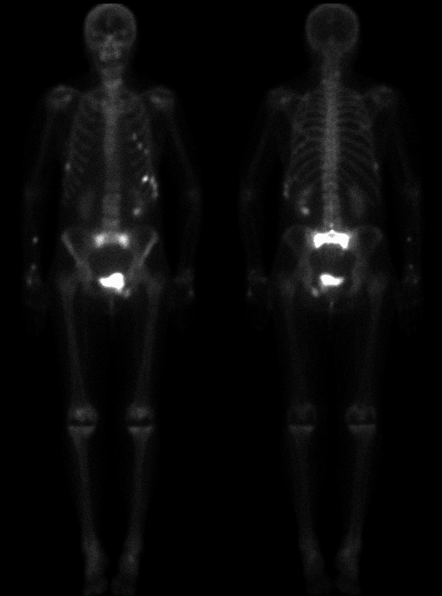
Anterior and posterior images are shown
View main image(bs) in a separate image viewer
View second image(bs).
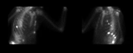
Spot views of the ribs.
View third image(mr).
T1 weighted image of the sacrum.
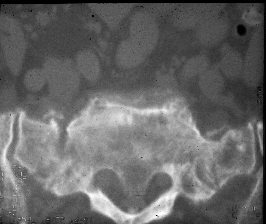
View fourth image(ct).
CT of sacrum.
Full history/Diagnosis is available below
Diagnosis: Sacral fracture, rib fractures
Full history:
81-year old woman with history
of multiple spontaneous rib fractures and presents
now with low back and sacral pain. Plain films of the
pelvis obtained revealed healing left inferior and
superior pubic rami fractures. MRI of the pelvis
obtained before bone scintigraphy demonstrated a
broad area of marrow replacement in the S1 and S2
segments of the sacrum with the differential diagnosis
lying between sacral insufficiency fracture and
neoplastic marrow infiltrative processes.
Findings:
The delayed whole body images from
bone scintigraphy demonstrate intense uptake of the
radiotracer in the sacral region involving both wings
of the sacrum as well as the interposed sacrum. This
finding, also known as the łHonda˛ sign (named for
the automakerąs emblem), is characteristic for sacral
insufficiency fracture. There is also focally increased
uptake in the region of the left superior and inferior
pubic rami correlating with the radiographic findings
of healing fractures. Also noted are multiple foci of
increased uptake in the left anterior ribs connecting
as two curved lines and are entirely consistent with
rib fractures.
Discussion:
Sacral insufficiency or stress
fractures occur commonly in patients who are
osteoporotic or who have undergone radiation
therapy. On bone scintigraphy, they commonly
appear as an łH˛ with vertical uptake in the sacral ala
and horizontal uptake in the sacrum. Fractures may
also be unilateral involving just one side of the sacrum
or may appear as a transverse band of increased
activity across the body of the sacrum.
Followup:
Shortly after bone scintigraphy, the
patient also had CT myelogram performed. Scans
through the sacrum confirmed the presence of
bilateral sacral insufficiency fractures.
ACR Codes and Keywords:
References and General Discussion of Bone Scintigraphy (Anatomic field:Skeletal System, Category:Effect of Trauma)
Search for similar cases.
Edit this case
Add comments about this case
Read comments about this case
Return to the Teaching File home page.
Case number: bs028
Copyright by Wash U MO