Case Author(s): John Lahorra, MD and Jerold Wallis, MD , 6/23/95 . Rating: #D2, #Q3
Diagnosis: Adult polycystic kidney disease
Brief history:
Lung cancer
Images:
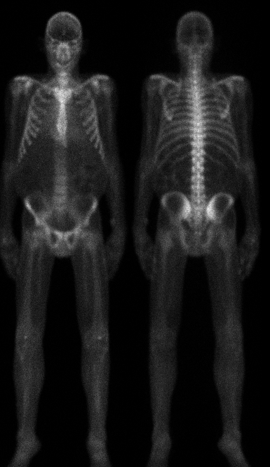
Whole body bone scan
View main image(bs) in a separate image viewer
Full history/Diagnosis is available below
Diagnosis: Adult polycystic kidney disease
Full history:
Patient with adult polycystic kidney disease on peritoneal dialysis, being evaulated after recent diagnosis of lung cancer.
Radiopharmaceutical:
Tc-99m MDP
Findings:
There is nonvisualization of the kidneys. The flanks are bulging slightly. There is no evidence of bony metastases.
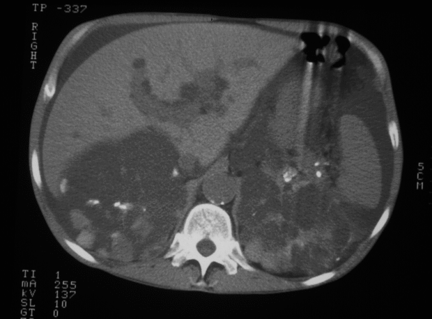
CT scan of the abdomen (see followup image) demonstrates bilateral, enlarged, cyst-replaced kidneys consistent with the patient's adult polycystic kidney disease.
View followup image(ct).
CT examination
Major teaching point(s):
In addition to viewing the bones, some attention to soft tissue uptake (or in this case, lack of soft tissue uptake) is useful.
ACR Codes and Keywords:
References and General Discussion of Bone Scintigraphy (Anatomic field:Genitourinary System, Category:Normal, Technique, Congenital Anomaly)
Search for similar cases.
Edit this case
Add comments about this case
Return to the Teaching File home page.
Case number: bs027
Copyright by Wash U MO