Case Author(s): virginia klaas , Henry Royal , 6-2-95 . Rating: #D2, #Q3
Diagnosis: Metastases, hypercalcemia, barium artifact
Brief history:
65 year old male with laryngeal carcinoma. Rule out metasases.
Images:

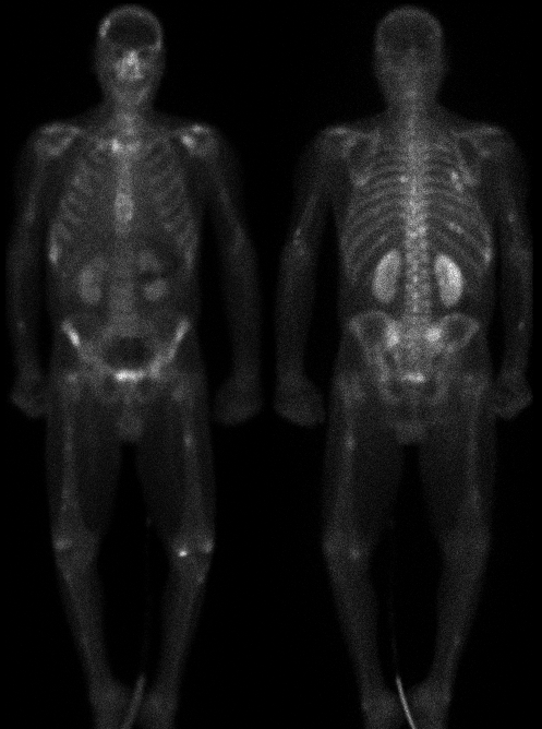
anterior and posterior whole body images.
View main image(bs) in a separate image viewer
Full history/Diagnosis is available below
Diagnosis: Metastases, hypercalcemia, barium artifact
Full history:
65-year old man with known laryngeal carcinoma, which
was resected approximately six months ago. The patient
is now referred to rule out bony metatases.
Findings:
Whole body bone scintigraphy demonstrates multiple foci
of increased uptake in the axial and appendicular skeleton,
which are most consistent with bone metastases.
Additionally, there is persistent increased
uptake in both kidneys. A curvilinear region of absent
uptake is seen over the left kidney on the anterior view
which correlates with residual barium from an upper GI
performed one day prior to bone scintigraphy.
Discussion:
Although extensive metastases is somewhat unusual in
patients with laryngeal carcinoma, computed tomography
demonstrated multiple pulmonary nodules, mediastinal
adenopathy, and bone destruction. Therefore, the
scintigraphic findings in this patient do correlate with
bone metastases. The differential diagnosis
of persistent increased uptake in both kidneys includes
dehydration, hypercalcemia, prior chemotherapy or
radiation therapy. The increased uptake in this patient was
known to be related to hypercalcemia, which was
documented by laboratory data.
Followup:
None
Major teaching point(s):
See discussion
ACR Codes and Keywords:
References and General Discussion of Bone Scintigraphy (Anatomic field:Skeletal System, Category:Neoplasm, Neoplastic-like condition)
Search for similar cases.
Edit this case
Add comments about this case
Read comments about this case
Return to the Teaching File home page.
Case number: bs025
Copyright by Wash U MO

