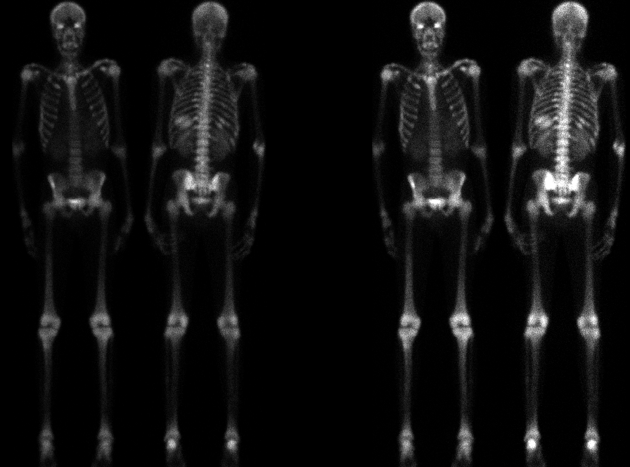

Anterior and posterior whole-body images, displayed at two different intensity settings.
View main image(bs) in a separate image viewer
View second image(iw). Another imaging study was obtained to help evaluate for infection.
View third image(bm). A final imaging study to supplement the second study. Why was it obtained?
Full history/Diagnosis is available below
A few weeks later, the patient again presented with pain at multiple sites, and an additional imaging was obtained.
In-111 labeled White Blood Cells
Tc-99m Sulfur Colloid
The second image set is an In-111 white blood cell study. Intense uptake is seen in the liver, with no uptake in the spleen. Activity is seen in the bone marrow, with areas of asymmetry (e.g. at the sacroiliac joints).
The third study is a Tc-99m sulfur colloid bone marrow study. It demonstrates the same sphenoid uptake as seen on bone scintigraphy, and the same sacroiliac joint pattern as seen on the In-111 white blood cell study.
On the white blood cell study, the previous splenic infarction results in the absence of uptake of labeled white blood cells in the spleen. The asymmetric uptake in the sacroiliac joints could represent infection or an asymmetric pattern of bone marrow expansion/infarction.
The bone marrow scintigraphy demonstrates an identical pattern of uptake as that seen on the white blood cell study, suggesting that no infection is present.
On the basis of the negative imaging study, antibiotics were discontinued. The fevers resolved, and the patient was subsequently discharged from the hospital.
2) The appearance of splenic infarction varies with scintigraphic tracer, as noted above.
3) The specificity of findings on In-111 white blood cell studies may be increased by additional imaging of the bone marrow using Tc-99m sulfur colloid. Areas of discordant uptake (greater on the In-111 white blood cell study than on the Tc-99m sulfur colloid study) favor infection. Equal activity on the two studies reflects the distribution of functioning bone marrow.
References and General Discussion of Bone Scintigraphy (Anatomic field:Skeletal System, Category:Other generalized systemic disorder)
Return to the Teaching File home page.