Case Author(s): Scott St. Amour, J. Walllis , 5/12/95 . Rating: #D2, #Q4
Diagnosis: Collimator contamination
Brief history:
Evaluate for metastatic disease
Images:
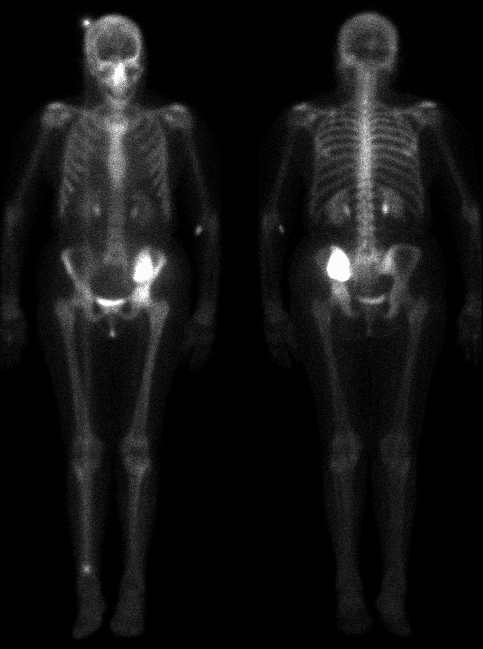
Anterior and posterior whole body images
View main image(bs) in a separate image viewer
Full history/Diagnosis is available below
Diagnosis: Collimator contamination
Full history:
The patient has lung cancer with a known pelvic metastasis.
Previous computed tomography studies have shown a large
expansile destrictive lesion in the left ilium, which
has been treated with radiation therapy.
Findings:
Markedly increased uptake is seen in the pelvis at the
site of the patients known tumor.
A linear area of
increased uptake is seen extending from the calvarium
to the right leg, most intense at the two ends of the
line, and apparent on the anterior view only.
Discussion:
The degree of increased uptake in the pelvis would be
compatible with either Paget's disease or tumor.
Previous CT examination suggests the latter.
The linear increased uptake is due to contamination of
the anterior detector, likely on the collimator. The
contamination is likely a Tc-99m tracer, since it was
apparent on the 140 kev bone imaging window without
excessive scatter or collimator penetration. A line
was generated due to the scanning motion whole
body camera. The
greater intensity at both ends is due to the fact that
the camera spends several minutes in stationary mode
at both ends of the body, in order to equalize time
over the body during the scan.
Major teaching point(s):
Contamination appears differently on whole body images
compared with spot views. In either case, it should
be recognized and not confused with bone lesions.
ACR Codes and Keywords:
- General ACR code: 49
- Skeletal System:
4.93 "ARTIFACT"
References and General Discussion of Bone Scintigraphy (Anatomic field:Skeletal System, Category:Other(Artifact))
Search for similar cases.
Edit this case
Add comments about this case
Read comments about this case
Return to the Teaching File home page.
Case number: bs022
Copyright by Wash U MO