Case Author(s): Vreeland, M.D./Wallis, M.D. , 2/11/95 . Rating: #D2, #Q4
Diagnosis: Radiation-induced hypoplasia
Brief history:
20 year old man on steroids with back/hip
pain
Images:
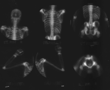
Spot views of the whole body
View main image(bs) in a separate image viewer
View second image(bs).
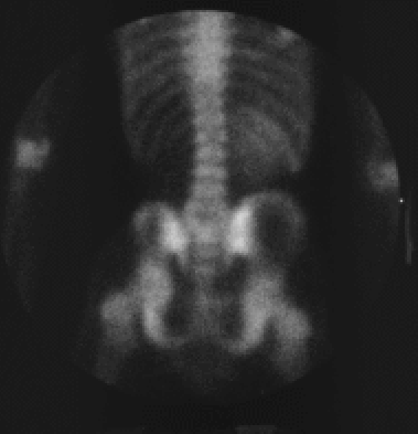
Enlargement of selected spot image.
View third image(xr).
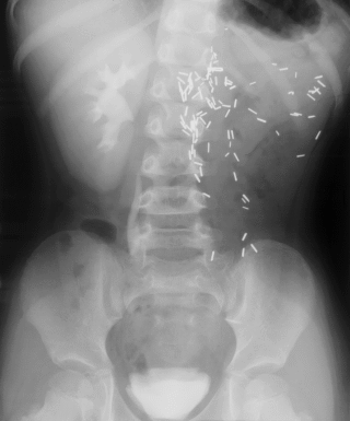
Plain films of the abdomen and pelvis.
Full history/Diagnosis is available below
Diagnosis: Radiation-induced hypoplasia
Full history:
20 year old man who is status post orthotopic heart
transplantation in 1987 for an idiopathic cardiomyopathy.
He also has a history of left-sided nephrectomy at the age
of three for a Wilm's tumor, followed by radiation therapy.
This study is being done to rule out avascular necrosis
of the hip.
Findings:
Bone Scintigraphy
(1) No scintigraphic evidence of avascular necrosis; the
anterior pelvis views (not shown) also demonstrated normal
uptake of tracer in the femoral heads.
(2) Hypoplastic left iliac wing and surgically absent left kidney,
consistent with the patient's history.
Radiograph of the Abdomen:
(1) Contrast is seen in the normal appearing remaining
right kidney and bladder.
(2) Multiple clips over the left abdomen confirm the
surgical absence of the left kidny.
(3) Radiographs also confirm the hypoplastic left iliac
wing.
Discussion:
Radiation damage to musculoskeletal tissues occurs initally
from the vascular inflamation and subsequent fibrosis.
Bone scintigraphy performed within
weeks of radiation therapy may show increased uptake in the
port area because of the inflamation. As weeks pass,
fibrotic changes cause decreased blood flow to the port
area, and subsequent bone scintigraphic evaluations would
show nonanatomic areas of decreased uptake corresponding
to the port. If high enough doses of radiation are used,
especially in young children, radiation-induced hypoplasia
will occur because of the cytotoxic effect to osteoblasts
which occur with doses greater than 1200 rads. The younger
the child, the more pronounced the asymmetry.
Approximately 2.5% of patients with Wilm's disease have
hemihypertrophy, which is usually noted on the side of the
body contralateral to the tumor. Hemihypertrophy should
not be restricted to a port area, and generally has activity
proportionate to bone mass.
Followup:
Conservative treatment.
ACR Codes and Keywords:
References and General Discussion of Bone Scintigraphy (Anatomic field:Skeletal System, Category:Misc)
Search for similar cases.
Edit this case
Add comments about this case
Read comments about this case
Return to the Teaching File home page.
Case number: bs018
Copyright by Wash U MO