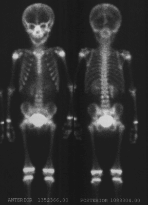
Anterior and posterior whole-body images from bone scintigraphy
View main image(bs) in a separate image viewer
View second image(mr). MRI images of the abdomen.
View third image(xr). Abdominal plain film showing the radiation port.
Full history/Diagnosis is available below
(1) Persistent, mildly increased activity is seen involving the pedicles of L3, without interval change compared to a prior bone scintigraphy performed at the time of development of pain (not included in this teaching file). No new foci of abnormally increased activity to otherwise suggest osseous metastatic disease are seen.
(2) Moderately increased activity is seen along the medial border of the right kidney representing an interval change compared to the prior examination. Therefore, in this patient with a history of recent radiation treatments, radiation nephritis is the most likely etiology.
MRI Examination:
(1) Enhancing, epidural mass extending from L2 - L4.
(2) Blastic involvement of the L3 verterbral body is unchanged.
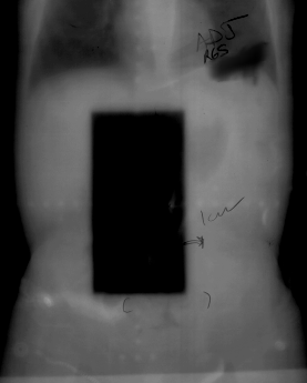
Abdominal X-ray:
The radiation port is seen.
(1) The threshold dose for development of nephritis is 2,300 rads
(2) May occur up to two years after treatment
(3) Clinically, the nephritis resembles a chronic glomerulonephritis with normal-to-small kidney(s) in the field of radiation. Parenchymal thickness in involved areas is also diminished.
(4) Associated with hypertension
(5) Histologically, one sees interstitial fibrosis, tubular atrophy, glomerular and arteriolar sclerosis.
View followup image(bs). Bone scintigraphy six months after radiation treatment shows resolution of the renal finding.
References and General Discussion of Bone Scintigraphy (Anatomic field:Genitourinary System, Category:Inflammation,Infection)
Return to the Teaching File home page.