Case Author(s): Hamid Latifi,MD, Jeff Dobkin,MD, Tom R. Miller,MD,PhD , 11/8/94 . Rating: #D3, #Q4
Diagnosis: Idiopathic Pulmonary Hemosiderosis
Brief history:
20 year-old woman with recurrent hemoptysis.
Images:

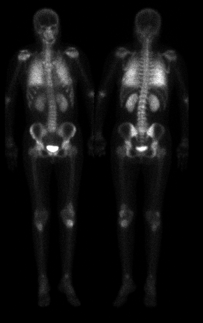
Above are anterior and posterior whole-body bone scintigraphy images.
View main image(bs) in a separate image viewer
Full history/Diagnosis is available below
Diagnosis: Idiopathic Pulmonary Hemosiderosis
Full history:
The patient is a 20 year-old woman with recurrent hemoptysis
and primary pulmonary hemosiderosis, nephrocalcinosis, mucormycosis
and chronic steroid use (for her pulmonary disease). She presented
with a 3-week history of back pain. A plain radiograph of the spine demonstrated questionable
wedging of the L1 vertebra. This study was requested to exclude fungal osteomyelitis of the spine.
Findings:
There is no evidence of vertebral osteomyelitis. Increased activity in both lungs and kidneys are noted, consistent with the patient's pulmonary hemosiderosis and nephrocalcinosis.
Increased uptake in proximal tibiae and left distal femur may be secondary to prior trauma, avascular necrosis, or other processes.
Discussion:
The main finding in this case is the diffusely increased MDP uptake in both lungs, in this patient with known pulmonary hemosiderosis.
Major teaching point(s):
This case demonstrates an example of bone-seeking radiopharmaceutical accumulation in non-osseous structures.
Differential Diagnosis List
1. Prior lung scintigraphy.
2. Alveolar microlithiasis.
3. Secondary pulmonaru hemosiderosis and ossification, e.g. mitral regurgitation.
ACR Codes and Keywords:
References and General Discussion of Bone Scintigraphy (Anatomic field:Lung, Mediastinum, and Pleura, Category:Misc)
Search for similar cases.
Edit this case
Add comments about this case
Read comments about this case
Return to the Teaching File home page.
Case number: bs012
Copyright by Wash U MO

