Case Author(s): Thomas H. Vreeland, M.D., Tom R. Miller, M.D., Ph.D. , 08/26/94 . Rating: #D2, #Q3
Diagnosis: Paget's Disease
Brief history:
54 year-old man with low back pain.
Images:

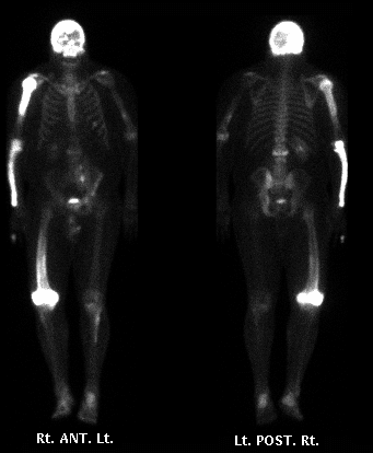
Bone Scintigraphy
View main image(bs) in a separate image viewer
Full history/Diagnosis is available below
Diagnosis: Paget's Disease
Full history:
54 year-old man with low back pain associated with an
injury in January, 1994. The pain has gradually worsened
over the past eight months.
Findings:
(1) Markedly increased bone uptake
involving the calvarium, right humerus, right forearm, the left tibia/fibula and
the distal right femur in a pattern characteristic of
Paget's Disease.
(2) Left pelvic kidney
(3) Increased uptake at L2
(4) Mildly increased activity in a lesion involving the
distal left humerus
(5) Subtle area of increased activity involving the left
superior pubic ramus and left ischium as well as well degenerative changes and costochondral junction activity
Discussion:
Paget's disease, along with fibrous dysplaysia, leads to the most intensely increased uptake seen by bone scintigraphy. Typically a large contiguous section of bone is involved. The activity at L2 may be due to Paget's disease with a suggestion of expansion of the vertebral body.
Differential Diagnosis List
1. Paget's disease.
2. Fibrous dysplasia.
3. Selected lesions could be due to metastases, infection or trauma (much less likely)
ACR Codes and Keywords:
- General ACR code: 48
- Skeletal System:
4.84 "PAGET DISEASE"
- Genitourinary System:
8.1435 "Inferior ectopia include: pelvic kidney, hypospadias exclude: ptosis (.133), ureterocele (.1454, .1455)"8.1435 "Inferior ectopia include: pelvic kidney, hypospadias exclude: ptosis (.133), ureterocele (.1454, .1455)"
References and General Discussion of Bone Scintigraphy (Anatomic field:Skeletal System, Category:Misc)
Search for similar cases.
Edit this case
Add comments about this case
Read comments about this case
Return to the Teaching File home page.
Case number: bs010
Copyright by Wash U MO

