Case Author(s): Vreeland, M.D. / Royal, M.D. , 08/24/94 . Rating: #D2, #Q3
Diagnosis: Enchondroma
Brief history:
35 year old woman with wrist and hand pain,
being evaluated with bone scintigraphy to
determine pattern of joint involvement.
Images:

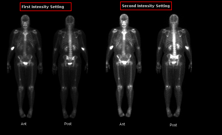
Whole Body Bone Scintigraphy
View main image(bs) in a separate image viewer
Full history/Diagnosis is available below
Diagnosis: Enchondroma
Full history:
This patient is a 35 year old woman with a 3 - 4 month
history of bilateral wrist and hand pain. In addition, she
has intermittent pain in the large joints of the knees and
shoulders. This examination is being done to determine
pattern of joint involvement and to rule out occult bony
lesions of the hands or wrists.
Findings:
(1) Mildly increased activity involving the metacarpal-
phalangeal joints of the second and third digits of both
hands; great toe of right foot; knees bilaterally; shoulders
bilaterally; and the tenth thoracic vertebral body in a
pattern consistent with arthritic changes. CXR (not included
in this case) demonstrated hypertrophic, degenerative changes
of the tenth thoracic vertebrate. Radiographs of the hands
(not included in this case) demonstrated minimal degenerative
changes of the third metacarpophalangeal joints bilaterally,
suggesting early degenerative changes or possible
hemochromatosis.
(2) Incidentally noted is focally, mildly increased
activity involving the distal third of the right femur.
Correlative radiographs of the femur demonstrate a lesion
most suggestive of enchondroma, with a bone infarct being
a less likely possibility. Clinically, the patient is
asymptomatic in this region.
(3) A region of extravasated radiopharmaceutical is
incidentially noted in the right antecubetal fossa.
Discussion:
This case was presented to demonstrate the scintigraphic
appearance of an enchondroma. Typically, enchondromas
have only mildly increased activity. Markedly increased
activity of enchondromas are most often associated with
pathologic fractures. If enchondromas are painless, they
are generally treated conservatively. Pain associated with
an enchondroma may represent malignant degeneration.
Enchondroma:
- Benign cartilaginous growth in bones
- M:F - 1:1
- Frequently multiple (Enchondromatosis or Ollier Disease)
- Often, in the small bones of the wrists and hands
- Can be found in diaphyseal or central regions of the
femur, tibia, humerus, radius, ulna, foot, or ribs.
- Generally, no cortical breakthrough or periosteal
reaction
- Malignant degeneration in long bone enchondromas can
occur in up to 15 -20% of cases, and are often associated
with pain.
Differential Diagnosis List
Differential Diagnosis for Enchondromas would include:
(1) Bone Infarct
(2) Chondrosarcoma
(3) Fibrous Dysplasia (rare in the hands)
(4) Unicameral bone cyst (rare in the hands)
(5) Epidermoid inclusion cyst
(6) Giant cell tumor of tendon sheath
ACR Codes and Keywords:
References and General Discussion of Bone Scintigraphy (Anatomic field:Skeletal System, Category:Neoplasm, Neoplastic-like condition)
Search for similar cases.
Edit this case
Add comments about this case
Read comments about this case
Return to the Teaching File home page.
Case number: bs009
Copyright by Wash U MO

