Case Author(s): Thomas H. Vreeland,MD , 07/16/94 . Rating: #D2, #Q4
Diagnosis: Inferior Vena Cava Obstruction
Brief history:
This patient is a 59 year old woman with a history of
uterine carcinoma, who is being evaluated to rule out
osseous metastatic disease.
Images:

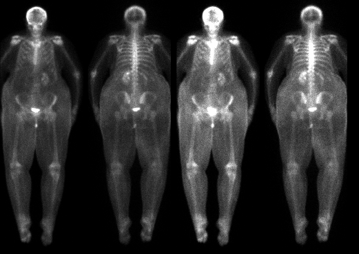
Anterior and Posterior views at different intensities
View main image(bs) in a separate image viewer
Full history/Diagnosis is available below
Diagnosis: Inferior Vena Cava Obstruction
Full history:
This patient is a 59 year old woman with a history of
uterine carcinoma, which was resected and treated with
radiation in 1991. The patient now complains of
progressive lower back pain. An MRI examination of the
lumbar spine performed on 7/15/94 demonstrated
inferior vena cava and bilateral common iliac
thrombosis. The patient was noted to have abnormal signal
in the sacrum (possible insufficiency
fracture) and involvement of the L3 vertebral body on
the MRI performed on 7/15/94. The patient is being evaluated to
rule out osseous metastatic disease.
Findings:
Bone Scintigraphy:(7/16/94)
(1) Marked soft tissue swelling of the abdomen and the
lower extremities
(2) Mildly increased activity in the third lumbar
vertebrate noted on anterior images
(3) No scintigraphic correlate for the sacral
abnormalities noted on the MRI from 7/15/94.
(4) Central area of photopenia involving the right
kidney in the region of the collecting system,
suggesting hydronephorsis
MRI Examination of the Lumbar Spine: (7/15/94)
(1) Possible invasion of L3 by adjacent soft tissue
mass
(2) Hydronephrosis of the right kidney
(3) No evidence of thecal sac or neural foramina
impingement
MRI Examination of Pelvis (7/14/94):
(1) Possible sacral insufficiency fracture
(2) IVC and bilateral common iliac vein thrombosis
Discussion:
(1) The marked soft tissue activity noted in the abdomen
and lower extremities, especially when compared to the
upper extremities, suggests the possibility of venous or
lymphatic obstruction.
(2) The scintigraphic findings involving L3 are nonspecific.
Degenerative changes would be the most common cause of such
scintigraphic findings.
(3) The findings involving the right kidney are relatively
nonspecific; however, in this setting, they are suspicious for
hydronephosis. Alternatively, a space-occupying lesion in
the collecting system (hematoma or tumor) would be included
in the differential diagnosis, in the proper clinical
setting.
Major teaching point(s):
Vascular or lymphatic obstruction should be suspected
when gross asymmetry of soft tissue activity is noted in one
extremity (or in half of the body in this case) when compared
to the rest of the body. The diagnosis of IVC obstruction
is often known or suspected in a patient prior to
scintigraphic examination; however, occasionally, this
diagnosis is first detected on scintigraphic examinations.
Differential Diagnosis List
(1) Vascular or lymphatic obstruction (intrinsic or extrinsic)
(2) Congenital
ACR Codes and Keywords:
References and General Discussion of Bone Scintigraphy (Anatomic field:Vascular and Lymphatic Systems, Category:Organ specific)
Search for similar cases.
Edit this case
Add comments about this case
Read comments about this case
Return to the Teaching File home page.
Case number: bs006
Copyright by Wash U MO

