Case Author(s): Jerry Wallis , 6/17/94 . Rating: #D1, #Q4
Diagnosis: Metatarsal fracture
Brief history:
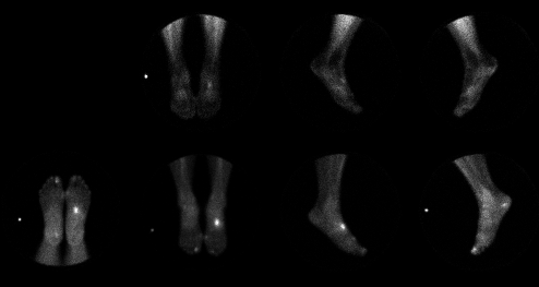
Dropped a steel drawer on her left foot 3 months ago. X-rays were negative (Upper row are immediate images and lower row are delayed images)
Images:

View main image(bs) in a separate image viewer
Full history/Diagnosis is available below
Diagnosis: Metatarsal fracture
Findings:
Mildly increase blood pool activity is seen (upper row) in the
mid tarsal region near the tarsal-metatarsal junction, with markedly increased delayed uptake
at the tarsal-metatarsal juction in the midfoot on the
delayed images
(lower row).
The increased uptake is somewhat difficult to localize, but is at
the junction of the 2nd or 3rd metatarsals and the
2nd or 3rd cuneiform, most likely in the proximal 3rd metatarsal.
Discussion:
This appearance is typical for a fracture, which may be due to increased
use or traumatic injury. Although less likely in this patient,
healing avascular necrosis and focal osteomyelitis (e.g. due to
a penetrating wound) could have a similar appearance.
On these 2 hour delay images, the limited uptake
in the normal bones makes it difficult
to localize the fracture precisely. If
needed, pinhole images or delayed images at 24 hours
would likely have
defined the bony anatomy more precisely; however these were
not done as clinical management would not have been affected.
Followup:
Followup radiographs were negative, but the bone scintigraphy
is sufficient for the diagnostis of fracture in this clinical
setting.
Major teaching point(s):
Bone scintigraphy is more sensitive than plain radiography
in detecting bone fractures.
ACR Codes and Keywords:
References and General Discussion of Bone Scintigraphy (Anatomic field:Skeletal System, Category:Effect of Trauma)
Search for similar cases.
Edit this case
Add comments about this case
Read comments about this case
Return to the Teaching File home page.
Case number: bs003
Copyright by Wash U MO

