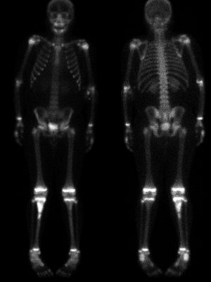
View main image(bs) in a separate image viewer
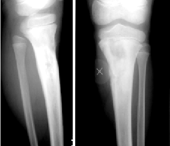
View second image(xr).
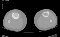
View third image(ct).
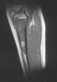
View fourth image(mr).
Full history/Diagnosis is available below
A mixed sclerotic/lytic lesion is seen that is felt to be most consistent with osteomyelitis or, less likely, osteosarcoma. A marker has been placed over the palpable mass.
Bone Scan:
There is markedly increased activity in the proximal 1/3 of the right tibia, with a second focus of intense activity slightly more distally. These findings are felt to be most consistent with an osteosarcoma with infectious a less likely possibility. No other abnormal foci are noted throughout the skeleton.
MRI:
A large soft tissue mass is seen within the proximal tibial metaphysis extending proximally into the epiphysis but not involving the knee joint. There is a "skip lesion" more distally in the tibial diaphysis. A break is seen in the medial cortex of the tibia with extension of the soft tissue mass which abuts but does not appear to evade the neurovascular bundle. All portions of the mass enhance with gadolinium.
CT:
There is a destructive lesion in the proximal right tibia with cortical breakthrough anteriorly and cortical tunneling medially. A small anterior soft tissue or fluid collection is seen subperiosteally. The appearance was felt to be atypical for osteosarcoma and more likely indicated osteomyelitis.
2. Bone scintigraphy is not the best way to distinguish osteomyelitis from osteosarcoma. However, in this example, the skip lesion (noted on both bone scintigraphy and MRI) strongly favored malignancy, especially osteosarcoma, over osteomyelitis which would otherwise have a similar appearance.
1. Osteosarcoma
2. Ewing's Sarcoma
3. Osteomyelitis
References and General Discussion of Bone Scintigraphy (Anatomic field:Skeletal System, Category:Neoplasm, Neoplastic-like condition)
Return to the Teaching File home page.