Case Author(s): Yonglin Pu, MD, PhD, Henry Royal, MD. , 04/16/03 . Rating: #D3, #Q4
Diagnosis: Extra-temporal Lobe Epilepsy
Brief history:
16 year old girl with medically intractable epilepsy. The patient has generalized tonic and clonic seizures on daily basis. Evaluate for epileptogenic zone.
Images:

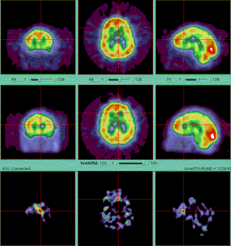
Perfusion Brain Imaging(Tomographic/Ictal and Interictal)
View main image(br) in a separate image viewer
View second image(pb).
Brain PET Imaging (Metabolism)
Full history/Diagnosis is available below
Diagnosis: Extra-temporal Lobe Epilepsy
Full history:
16-year-old female with an intractable seizure disorder. She has generalized tonic clonic seizures on a daily basis. She reports no loss of consciousness. The patient is currently on three anti seizure
medications. There is no history of trauma, febrile illness, or seizure disorder within the family. There are no lateralizing symptoms as reported by the patient or her mother.
Radiopharmaceutical:
12.0 mCi Tc-99m bicisate i.v and 15.4 mCi Tc-99m bicisate i.v. for ictal and interictal perfusion brain SPECT imagings. 7.8 mCi F-18 Fluorodeoxyglucose i.v. for metabolism brain PET imaging.
Findings:
An ictal examination was first performed during continuous video and electroencephalographic monitoring. Tc-99m bicisate was injected intravenously approximately 15 seconds after the electroencephalographic, and clinical onset of the seizures, which were characterized by tonic and clonic activity. Standard tomographic imaging (SPECT) of the brain was performed 1.5 hours after injection of the tracer.
For comparison with the ictal study, an interictal study was performed next day with same imaging protocol. At the time of this interictal study, the patient had been seizure free for approximately 24 hours.
The subtraction SPECT images demonstrate a discrete focus of increased activity within the anterior cingulate gyrus on the left. In addition, there is a less intense focus in the region of the left hippocampus. On the interictal SPECT, there is mild hypoperfusion of the anterior cingulate gyrus on the left as well, as correlated with the subtraction images. Specifically, the focal abnormality in the left hippocampus correlates with the mesial hypometabolism seen in the left temporal lobe on the FDG PET images performed next day.
Discussion:
Technetium 99m N,Ní-1,2-ethylenediylbis-L-cysteine diethylester(99m-Tc ECD, which is also known as Neurolite, is a lipophilic agent that crosses the blood-brain barrier with a rapid first-pass uptake. Once in the brain it is metabolized into a polar complex that retained in the brain. The brain uptake of the Neurolite is proportional to the cerebral blood flow (CBF) at the time of the injection of the tracer. Previous research has demonstrated good correlation of the regional cerebral blood flow and the uptake of Neurolite.
During a seizure, the regional CBF increases, and by measuring regional CBF during both ictal and interictal periods, we can demonstrate regional CBF changes due to seizure activity. It is important to inject the cerebral blood flow tracer within seconds of the start of the seizure to most accurately identify the epileptogenic zone in the brain. SPECT imaging with Neurolite can be used to estimate the localization of a epileptogenic zone and pre-surgical evaluation of epilepsy patients.
Ictal FDG brain imaging is not helpful identifying the epileptogenic focus because the slow uptake of FDG does not permit accurate localization of the epileptogenic focus. Interictal FDG brain imaging is more accurate than interictal SPECT brain blood flow imaging in identifying the epileptogenic focus.
References:
1. Saha GB, Maclntyre WJ and Go RT. Seminars in Nuclear Medicine 1994;XXIV(4):324-349.
Followup:
Patient had multiple subpial transection of motor cortex down to the level of the cingulate gyrus above the corpus callosum. She has been seizure free since the surgery.
Major teaching point(s):
Blood flow imaging with neurolite using SPECT technique is useful in the localization of the epileptogenic zone.
ACR Codes and Keywords:
References and General Discussion of Brain Scintigraphy (Anatomic field:Skull and Contents, Category:Misc)
Search for similar cases.
Edit this case
Add comments about this case
Return to the Teaching File home page.
Case number: br004
Copyright by Wash U MO

