Case Author(s): Jeffrey Yu, M.D. and Keith Fischer, M.D. , 6/29/99 and 6/30/99 . Rating: #D3, #Q4
Diagnosis: Positive Balloon Occlusion Test
Brief history:
45 Year Old Female presenting with headaches.
Images:

Axial brain SPECT images during total balloon occlusion and at baseline.
View main image(br) in a separate image viewer
View second image(ct).
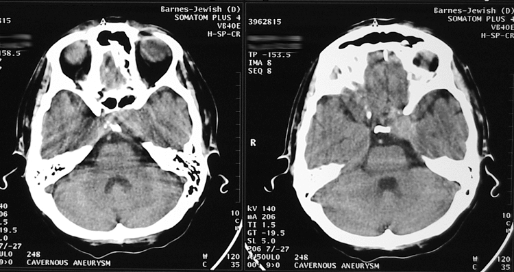
Selected non-contrast CT images of the head.
View third image(ct).
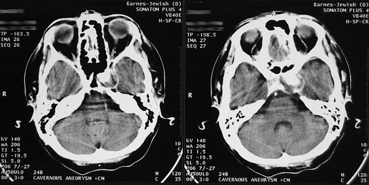
Selected contrast-enhanced CT images of the head.
View fourth image(an).
PA and lateral DSA images.
Full history/Diagnosis is available below
Diagnosis: Positive Balloon Occlusion Test
Full history:
45 year old woman who presented with headache and left cranial neuropathy.
Radiopharmaceutical:
Tc-99m Bicisate
Findings:
CT scan of the head reveals an area of increased attenuation in the left cavernous sinus region which enhances with contrast administration. A cavernous sinus aneurysm was suspected and an angiogram was performed which confirmed a left cavernous sinus aneurysm. Total balloon occlusion of that side was performed to evaluate for sufficient collateral circulation for possible vessel sacrifice. Tc-99m bicisate was injected during total balloon occlusion and SPECT imaging was performed. This demonstrated decreased radiotracer activity in the left frontal lobe extending inferiorly to the region of the Sylvian fissure. Quantitatively, the perfusion was about 20% less than the comparable region on the right. A baseline study was then performed the following day which demonstrated symmetric perfusion of both right and left cerebral hemispheres.
Discussion:
Tc-99m Bicisate can be injected under the desired conditions and imaged optimally up to 1.5 hours later. This allows for a great deal of flexibility in adapting the study to answer a wide variety of clinical problems. In this case, the clinical question was one of adequacy of collateral blood flow if the ipsilateral carotid artery was sacrificed. Tc-99m Bicisate answers this question as it can be injected during angiographic total balloon occlusion of the ipsilateral carotid, as in this case. Thus, the tracer distributes according to blood flow at the time of injection. In this case, it demonstrated the relatively decreased flow on the left that occurred during balloon occlusion of that side.
With the occlusion study alone, one cannot be certain if the changes seen are related to the occlusion or simply represent underlying atherosclerotic disease or the sequellae of previous infection or infarction. Thus, a baseline study was performed the next day. This involved re-injection of Tc-99m Bicisate without balloon occlusion. If the difference in perfusion is not present on the baseline study, then it is likely to have been created by the balloon occlusion. One can then quantify the percent decrease in activity by observing the change in color of a given region when using the segmented PET color scale.
Followup:
The patient had not had clinical symptoms during the occlusion but there was insufficient collateral flow angiographically when the ICA was occluded distal to the internal maxillary artery on that side. With the balloon positioned more proximally thereby allowing collateral flow from the internal maxillary artery, there was sufficient flow angiographically.
Thus, the SPECT tipped the scales and the patient went for middle meningeal to middle cerebral artery collateralization prior to internal carotid artery sacrifice.
Major teaching point(s):
Importance of obtaining a baseline study when abnormalities are seen on a post-occlusion brain SPECT
Differential Diagnosis List
Meningioma,
Cavernous Sinus Aneurysm
ACR Codes and Keywords:
References and General Discussion of Brain Scintigraphy (Anatomic field:Skull and Contents, Category:Organ specific)
Search for similar cases.
Edit this case
Add comments about this case
Read comments about this case
Return to the Teaching File home page.
Case number: br003
Copyright by Wash U MO