Case Author(s): J. Philip Moyers, M.D., Tom R. Miller, M.D., Ph.D. , 7/28/95 . Rating: #D2, #Q4
Diagnosis: Normal brain perfusion study
Brief history:
Evaluate for brain death.
Images:
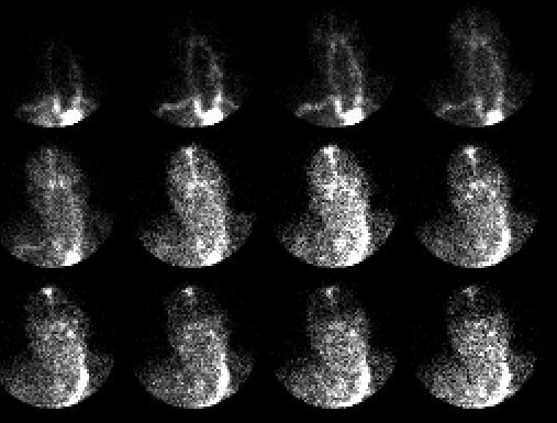
Anterior perfusion images of the head
View main image(br) in a separate image viewer
Full history/Diagnosis is available below
Diagnosis: Normal brain perfusion study
Full history:
This is a 76-year old man who
underwent surgery yesterday for repair of a
thoracoabdominal aneurysm. He now has nonreactive
pupils, and brain scintigraphy is requested to
evaluate for brain death.
Radiopharmaceutical:
Tc-99m DTPA
Findings:
This is a normal study with prompt,
symmetric perfusion of both cerebral
hemispheres. These two-second anterior images
obtained after bolus injection in an antecubital fossa
show perfusion of both common carotid arteries
followed by passage of the bolus through the circle of
Willis. On the third image, activity is seen in the
anterior and middle cerebral arteries, followed by a
capillary phase and, beginning in the fifth image, the
venous sinuses.
Discussion:
A prolonged bolus injection may
lead to no-diagnostic perfusion images where clear
separation of the phases of perfusion through the
carotid arteries and intracranial vascular system is
not seen. A similar appearance can be seen in
patients with congestive heart failure who have a
prolonged central circulation time. Localized reduced
perfusion can be caused by occlusive disease,
neoplasm, hematoma, a cystic lesion, or edema.
Localized increased perfusion can be caused by
arterial venous malformation, tumor, vascular
metastases, meningioma, or inflammatory lesion. A
dose of 15-25 mCi Tc-99m DTPA is usually used for
this study. A diagnosis of "no effective cerebral
perfusion" is made when there is a good bolus,
assessed by evaluation of the common carotid arteries,
with no flow through the circle of Willis, anterior and
middle cerebral arteries, and no capillary phase.
Faint venous sinus activity does not preclude this
interpretation. This study is useful in making the
diagnosis of brain death when supporting clinical data
are present.
References: 1) Mettler
FA. Essentials of Nuclear Medicine Imaging. 1991,
3rd edition. 2) Datz FL. Handbook of Nuclear
Medicine, Mosby Yearbook Publishers, 1993, 2nd
edition.
Major teaching point(s):
Appreciation of the
scintigraphic appearance of normal brain perfusion is
useful in accurate evaluation of brain death.
ACR Codes and Keywords:
References and General Discussion of Brain Scintigraphy (Anatomic field:Skull and Contents, Category:Normal, Technique, Congenital Anomaly)
Search for similar cases.
Edit this case
Add comments about this case
Read comments about this case
Return to the Teaching File home page.
Case number: br001
Copyright by Wash U MO